Ceon Ramon, Paolo Garguilo, Egill A Fridgeirsson, Jens Haueisen
{"title":"脑电图模拟人脑模型中硬脑膜层包涵对头皮电位变化及空间平滑效应的影响。","authors":"Ceon Ramon, Paolo Garguilo, Egill A Fridgeirsson, Jens Haueisen","doi":"10.3389/fneng.2014.00032","DOIUrl":null,"url":null,"abstract":"<p><p>The dura layer which covers the brain is less conductive than the CSF (cerebrospinal fluid) and also more conductive than the skull bone. This could significantly influence the flow of volume currents from cortex to the scalp surface which will also change the magnitude and spatial profiles of scalp potentials. This was examined with a 3-D finite element method (FEM) model of an adult subject constructed from 192 segmented axial magnetic resonance (MR) slices with 256×256 pixel resolution. The voxel resolution was 1×1×1 mm. The model included the dura layer. In addition, other major tissues were also identified. The electrical conductivities of various tissues were obtained from the literature. The conductivities of dura and CSF were 0.001 S/m and 0.06 S/m, respectively. The electrical activity of the cortex was represented by 144,000 distributed dipolar sources with orientations normal to the local cortical surface. The dipolar intensity was in the range of 0.0-0.4 mA meter with a uniform random distribution. Scalp potentials were simulated for two head models with an adaptive finite element solver. One model had the dura layer and in the other model, dura layer was replaced with the CSF. Spatial contour plots of potentials on the cortical surface, dural surface and the scalp surface were made. With the inclusion of the dura layer, scalp potentials decrease by about 20%. The contours of gyri and sulci structures were visible in the spatial profiles of the cortical potentials which were smoothed out on the dural surface and were not visible on the scalp surface. These results suggest that dura layer should be included for an accurate modeling of scalp and cortical potentials. </p>","PeriodicalId":73093,"journal":{"name":"Frontiers in neuroengineering","volume":" ","pages":"32"},"PeriodicalIF":0.0000,"publicationDate":"2014-08-05","publicationTypes":"Journal Article","fieldsOfStudy":null,"isOpenAccess":false,"openAccessPdf":"https://sci-hub-pdf.com/10.3389/fneng.2014.00032","citationCount":"26","resultStr":"{\"title\":\"Changes in scalp potentials and spatial smoothing effects of inclusion of dura layer in human head models for EEG simulations.\",\"authors\":\"Ceon Ramon, Paolo Garguilo, Egill A Fridgeirsson, Jens Haueisen\",\"doi\":\"10.3389/fneng.2014.00032\",\"DOIUrl\":null,\"url\":null,\"abstract\":\"<p><p>The dura layer which covers the brain is less conductive than the CSF (cerebrospinal fluid) and also more conductive than the skull bone. This could significantly influence the flow of volume currents from cortex to the scalp surface which will also change the magnitude and spatial profiles of scalp potentials. This was examined with a 3-D finite element method (FEM) model of an adult subject constructed from 192 segmented axial magnetic resonance (MR) slices with 256×256 pixel resolution. The voxel resolution was 1×1×1 mm. The model included the dura layer. In addition, other major tissues were also identified. The electrical conductivities of various tissues were obtained from the literature. The conductivities of dura and CSF were 0.001 S/m and 0.06 S/m, respectively. The electrical activity of the cortex was represented by 144,000 distributed dipolar sources with orientations normal to the local cortical surface. The dipolar intensity was in the range of 0.0-0.4 mA meter with a uniform random distribution. Scalp potentials were simulated for two head models with an adaptive finite element solver. One model had the dura layer and in the other model, dura layer was replaced with the CSF. Spatial contour plots of potentials on the cortical surface, dural surface and the scalp surface were made. With the inclusion of the dura layer, scalp potentials decrease by about 20%. The contours of gyri and sulci structures were visible in the spatial profiles of the cortical potentials which were smoothed out on the dural surface and were not visible on the scalp surface. These results suggest that dura layer should be included for an accurate modeling of scalp and cortical potentials. </p>\",\"PeriodicalId\":73093,\"journal\":{\"name\":\"Frontiers in neuroengineering\",\"volume\":\" \",\"pages\":\"32\"},\"PeriodicalIF\":0.0000,\"publicationDate\":\"2014-08-05\",\"publicationTypes\":\"Journal Article\",\"fieldsOfStudy\":null,\"isOpenAccess\":false,\"openAccessPdf\":\"https://sci-hub-pdf.com/10.3389/fneng.2014.00032\",\"citationCount\":\"26\",\"resultStr\":null,\"platform\":\"Semanticscholar\",\"paperid\":null,\"PeriodicalName\":\"Frontiers in neuroengineering\",\"FirstCategoryId\":\"1085\",\"ListUrlMain\":\"https://doi.org/10.3389/fneng.2014.00032\",\"RegionNum\":0,\"RegionCategory\":null,\"ArticlePicture\":[],\"TitleCN\":null,\"AbstractTextCN\":null,\"PMCID\":null,\"EPubDate\":\"2014/1/1 0:00:00\",\"PubModel\":\"eCollection\",\"JCR\":\"\",\"JCRName\":\"\",\"Score\":null,\"Total\":0}","platform":"Semanticscholar","paperid":null,"PeriodicalName":"Frontiers in neuroengineering","FirstCategoryId":"1085","ListUrlMain":"https://doi.org/10.3389/fneng.2014.00032","RegionNum":0,"RegionCategory":null,"ArticlePicture":[],"TitleCN":null,"AbstractTextCN":null,"PMCID":null,"EPubDate":"2014/1/1 0:00:00","PubModel":"eCollection","JCR":"","JCRName":"","Score":null,"Total":0}
引用次数: 26
摘要
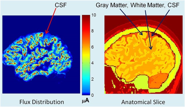
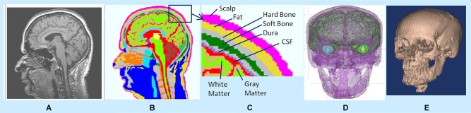
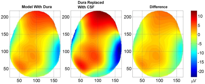
覆盖大脑的硬脑膜层的导电性不如脑脊液(CSF),但也比颅骨导电性好。这可能会显著影响从皮层到头皮表面的体积电流的流动,这也会改变头皮电位的大小和空间分布。这是通过成人受试者的三维有限元方法(FEM)模型来验证的,该模型由192个轴向磁共振(MR)切片以256×256像素分辨率构建而成。体素分辨率为1×1×1 mm。该模型包括硬脑膜层。此外,还鉴定了其他主要组织。从文献中得到了各种组织的电导率。硬脑膜和脑脊液电导率分别为0.001 S/m和0.06 S/m。皮层的电活动由144,000个分布的偶极源表示,这些偶极源的方向与局部皮层表面垂直。偶极子强度在0.0 ~ 0.4 mA m范围内,呈均匀随机分布。用自适应有限元求解器模拟了两种头部模型的头皮电位。一个模型有硬脑膜层,另一个模型用脑脊液代替硬脑膜层。绘制皮层表面、硬脑膜表面和头皮表面电位的空间等高线图。随着硬脑膜层的加入,头皮电位降低约20%。脑回和脑沟结构的轮廓在硬脑膜表面平滑的皮质电位空间剖面中可见,而在头皮表面不可见。这些结果表明,硬脑膜层应该包括在准确的头皮和皮质电位模型中。
Changes in scalp potentials and spatial smoothing effects of inclusion of dura layer in human head models for EEG simulations.
The dura layer which covers the brain is less conductive than the CSF (cerebrospinal fluid) and also more conductive than the skull bone. This could significantly influence the flow of volume currents from cortex to the scalp surface which will also change the magnitude and spatial profiles of scalp potentials. This was examined with a 3-D finite element method (FEM) model of an adult subject constructed from 192 segmented axial magnetic resonance (MR) slices with 256×256 pixel resolution. The voxel resolution was 1×1×1 mm. The model included the dura layer. In addition, other major tissues were also identified. The electrical conductivities of various tissues were obtained from the literature. The conductivities of dura and CSF were 0.001 S/m and 0.06 S/m, respectively. The electrical activity of the cortex was represented by 144,000 distributed dipolar sources with orientations normal to the local cortical surface. The dipolar intensity was in the range of 0.0-0.4 mA meter with a uniform random distribution. Scalp potentials were simulated for two head models with an adaptive finite element solver. One model had the dura layer and in the other model, dura layer was replaced with the CSF. Spatial contour plots of potentials on the cortical surface, dural surface and the scalp surface were made. With the inclusion of the dura layer, scalp potentials decrease by about 20%. The contours of gyri and sulci structures were visible in the spatial profiles of the cortical potentials which were smoothed out on the dural surface and were not visible on the scalp surface. These results suggest that dura layer should be included for an accurate modeling of scalp and cortical potentials.


 分享
分享

 求助内容:
求助内容: 应助结果提醒方式:
 扫码关注我们
