Francesca Ravanetti, Carlo Galli, Edoardo Manfredi, Anna Maria Cantoni, Edoardo Scarpa, Guido Maria Macaluso, Antonio Cacchioli
{"title":"D-(+)棉子糖修饰壳聚糖基支架用于软骨修复的体内研究。","authors":"Francesca Ravanetti, Carlo Galli, Edoardo Manfredi, Anna Maria Cantoni, Edoardo Scarpa, Guido Maria Macaluso, Antonio Cacchioli","doi":"10.1186/s12952-014-0021-5","DOIUrl":null,"url":null,"abstract":"<p><strong>Background: </strong>Osteochondral defects significantly affect patients' quality of life and represent challenging tissue lesions, because of the poor regenerative capacity of cartilage. Tissue engineering has long sought to promote cartilage repair, by employing artificial scaffolds to enhance cell capacity to deposit new cartilage. An ideal biomaterial should closely mimic the natural environment of the tissue, to promote scaffold colonization, cell differentiation and the maintenance of a differentiated cellular phenotype. The present study evaluated chitosan scaffolds enriched with D-(+) raffinose in osteochondral defects in rabbits. Cartilage defects were created in distal femurs, both on the condyle and on the trochlea, and were left untreated or received a chitosan scaffold. The animals were sacrificed after 2 or 4 weeks, and samples were analysed microscopically.</p><p><strong>Results: </strong>The retrieved implants were surrounded by a fibrous capsule and contained a noticeable inflammatory infiltrate. No hyaline cartilage was formed in the defects. Although defect closure reached approximately 100% in the control group after 4 weeks, defects did not completely heal when filled with chitosan. In these samples, the lesion contained granulation tissue at 2 weeks, which was then replaced by fibrous connective tissue by week 4. Noteworthy, chitosan never appeared to be integrated in the surrounding cartilage.</p><p><strong>Conclusions: </strong>In conclusion, the present study highlights the limits of D-(+) raffinose-enriched chitosan for cartilage regeneration and offers useful information for further development of this material for tissue repair.</p>","PeriodicalId":73849,"journal":{"name":"Journal of negative results in biomedicine","volume":"14 ","pages":"2"},"PeriodicalIF":0.0000,"publicationDate":"2015-01-14","publicationTypes":"Journal Article","fieldsOfStudy":null,"isOpenAccess":false,"openAccessPdf":"https://sci-hub-pdf.com/10.1186/s12952-014-0021-5","citationCount":"11","resultStr":"{\"title\":\"Chitosan-based scaffold modified with D-(+) raffinose for cartilage repair: an in vivo study.\",\"authors\":\"Francesca Ravanetti, Carlo Galli, Edoardo Manfredi, Anna Maria Cantoni, Edoardo Scarpa, Guido Maria Macaluso, Antonio Cacchioli\",\"doi\":\"10.1186/s12952-014-0021-5\",\"DOIUrl\":null,\"url\":null,\"abstract\":\"<p><strong>Background: </strong>Osteochondral defects significantly affect patients' quality of life and represent challenging tissue lesions, because of the poor regenerative capacity of cartilage. Tissue engineering has long sought to promote cartilage repair, by employing artificial scaffolds to enhance cell capacity to deposit new cartilage. An ideal biomaterial should closely mimic the natural environment of the tissue, to promote scaffold colonization, cell differentiation and the maintenance of a differentiated cellular phenotype. The present study evaluated chitosan scaffolds enriched with D-(+) raffinose in osteochondral defects in rabbits. Cartilage defects were created in distal femurs, both on the condyle and on the trochlea, and were left untreated or received a chitosan scaffold. The animals were sacrificed after 2 or 4 weeks, and samples were analysed microscopically.</p><p><strong>Results: </strong>The retrieved implants were surrounded by a fibrous capsule and contained a noticeable inflammatory infiltrate. No hyaline cartilage was formed in the defects. Although defect closure reached approximately 100% in the control group after 4 weeks, defects did not completely heal when filled with chitosan. In these samples, the lesion contained granulation tissue at 2 weeks, which was then replaced by fibrous connective tissue by week 4. Noteworthy, chitosan never appeared to be integrated in the surrounding cartilage.</p><p><strong>Conclusions: </strong>In conclusion, the present study highlights the limits of D-(+) raffinose-enriched chitosan for cartilage regeneration and offers useful information for further development of this material for tissue repair.</p>\",\"PeriodicalId\":73849,\"journal\":{\"name\":\"Journal of negative results in biomedicine\",\"volume\":\"14 \",\"pages\":\"2\"},\"PeriodicalIF\":0.0000,\"publicationDate\":\"2015-01-14\",\"publicationTypes\":\"Journal Article\",\"fieldsOfStudy\":null,\"isOpenAccess\":false,\"openAccessPdf\":\"https://sci-hub-pdf.com/10.1186/s12952-014-0021-5\",\"citationCount\":\"11\",\"resultStr\":null,\"platform\":\"Semanticscholar\",\"paperid\":null,\"PeriodicalName\":\"Journal of negative results in biomedicine\",\"FirstCategoryId\":\"1085\",\"ListUrlMain\":\"https://doi.org/10.1186/s12952-014-0021-5\",\"RegionNum\":0,\"RegionCategory\":null,\"ArticlePicture\":[],\"TitleCN\":null,\"AbstractTextCN\":null,\"PMCID\":null,\"EPubDate\":\"\",\"PubModel\":\"\",\"JCR\":\"\",\"JCRName\":\"\",\"Score\":null,\"Total\":0}","platform":"Semanticscholar","paperid":null,"PeriodicalName":"Journal of negative results in biomedicine","FirstCategoryId":"1085","ListUrlMain":"https://doi.org/10.1186/s12952-014-0021-5","RegionNum":0,"RegionCategory":null,"ArticlePicture":[],"TitleCN":null,"AbstractTextCN":null,"PMCID":null,"EPubDate":"","PubModel":"","JCR":"","JCRName":"","Score":null,"Total":0}
Chitosan-based scaffold modified with D-(+) raffinose for cartilage repair: an in vivo study.
Background: Osteochondral defects significantly affect patients' quality of life and represent challenging tissue lesions, because of the poor regenerative capacity of cartilage. Tissue engineering has long sought to promote cartilage repair, by employing artificial scaffolds to enhance cell capacity to deposit new cartilage. An ideal biomaterial should closely mimic the natural environment of the tissue, to promote scaffold colonization, cell differentiation and the maintenance of a differentiated cellular phenotype. The present study evaluated chitosan scaffolds enriched with D-(+) raffinose in osteochondral defects in rabbits. Cartilage defects were created in distal femurs, both on the condyle and on the trochlea, and were left untreated or received a chitosan scaffold. The animals were sacrificed after 2 or 4 weeks, and samples were analysed microscopically.
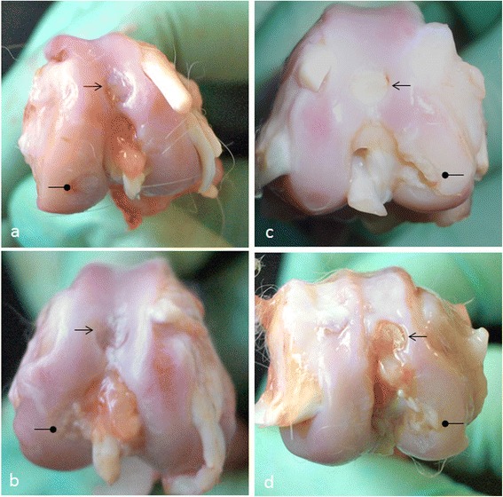
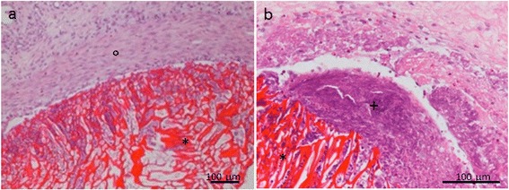
Results: The retrieved implants were surrounded by a fibrous capsule and contained a noticeable inflammatory infiltrate. No hyaline cartilage was formed in the defects. Although defect closure reached approximately 100% in the control group after 4 weeks, defects did not completely heal when filled with chitosan. In these samples, the lesion contained granulation tissue at 2 weeks, which was then replaced by fibrous connective tissue by week 4. Noteworthy, chitosan never appeared to be integrated in the surrounding cartilage.
Conclusions: In conclusion, the present study highlights the limits of D-(+) raffinose-enriched chitosan for cartilage regeneration and offers useful information for further development of this material for tissue repair.


 分享
分享

 求助内容:
求助内容: 应助结果提醒方式:
 扫码关注我们
