Nicole Rubin, Ali Darehzereshki, Saverio Bellusci, Vesa Kaartinen, Ching Ling Lien
{"title":"在新生小鼠心脏修复过程中,FGF10 信号可促进心外膜细胞扩张","authors":"Nicole Rubin, Ali Darehzereshki, Saverio Bellusci, Vesa Kaartinen, Ching Ling Lien","doi":"10.4172/2329-9517.1000101","DOIUrl":null,"url":null,"abstract":"<p><p>Unlike zebrafish and newt hearts, mammalian hearts have limited capacity to regenerate. Upon injury or disease, the adult mammalian hearts form a fibrotic scar. Recently, it was shown that neonatal mouse hearts can regenerate similarly to adult zebrafish hearts. However, this capacity quickly decreases after postnatal day 7 (P7). Understanding the molecular mechanisms underlying neonatal heart regeneration might lead to therapeutic approaches for regenerating adult mammalian hearts. In this study, we utilized an inducible transgenic mouse model to determine the effects of FGF10 growth factor over expression on neonatal mouse heart regeneration/repair. Over expression of FGF10 in myocardium enhanced the expansion of Wt1 positive epicardial cells at 21 days after heart injury through increased proliferation. However, this expansion of epicardial cells did not lead to increased epithelial-to-mesenchymal transition or affect fibroblast formation or fibrosis, as seen by vimentin expression, after heart injury. Furthermore, neither continuous nor transient expression of FGF10 did not affect scar thickness or length after heart injury in neonatal hearts. Our results suggest that FGF10 can regulate epicardial cell expansion of neonatal mouse hearts after injury; however, FGF10 alone is not sufficient to cause beneficial effects on heart repair.</p>","PeriodicalId":73638,"journal":{"name":"Journal of cardiovascular diseases & diagnosis","volume":"1 1","pages":""},"PeriodicalIF":0.0000,"publicationDate":"2013-03-01","publicationTypes":"Journal Article","fieldsOfStudy":null,"isOpenAccess":false,"openAccessPdf":"https://ftp.ncbi.nlm.nih.gov/pub/pmc/oa_pdf/ac/b9/nihms680985.PMC4407283.pdf","citationCount":"0","resultStr":"{\"title\":\"<i>FGF10</i> Signaling Enhances Epicardial Cell Expansion during Neonatal Mouse Heart Repair.\",\"authors\":\"Nicole Rubin, Ali Darehzereshki, Saverio Bellusci, Vesa Kaartinen, Ching Ling Lien\",\"doi\":\"10.4172/2329-9517.1000101\",\"DOIUrl\":null,\"url\":null,\"abstract\":\"<p><p>Unlike zebrafish and newt hearts, mammalian hearts have limited capacity to regenerate. Upon injury or disease, the adult mammalian hearts form a fibrotic scar. Recently, it was shown that neonatal mouse hearts can regenerate similarly to adult zebrafish hearts. However, this capacity quickly decreases after postnatal day 7 (P7). Understanding the molecular mechanisms underlying neonatal heart regeneration might lead to therapeutic approaches for regenerating adult mammalian hearts. In this study, we utilized an inducible transgenic mouse model to determine the effects of FGF10 growth factor over expression on neonatal mouse heart regeneration/repair. Over expression of FGF10 in myocardium enhanced the expansion of Wt1 positive epicardial cells at 21 days after heart injury through increased proliferation. However, this expansion of epicardial cells did not lead to increased epithelial-to-mesenchymal transition or affect fibroblast formation or fibrosis, as seen by vimentin expression, after heart injury. Furthermore, neither continuous nor transient expression of FGF10 did not affect scar thickness or length after heart injury in neonatal hearts. Our results suggest that FGF10 can regulate epicardial cell expansion of neonatal mouse hearts after injury; however, FGF10 alone is not sufficient to cause beneficial effects on heart repair.</p>\",\"PeriodicalId\":73638,\"journal\":{\"name\":\"Journal of cardiovascular diseases & diagnosis\",\"volume\":\"1 1\",\"pages\":\"\"},\"PeriodicalIF\":0.0000,\"publicationDate\":\"2013-03-01\",\"publicationTypes\":\"Journal Article\",\"fieldsOfStudy\":null,\"isOpenAccess\":false,\"openAccessPdf\":\"https://ftp.ncbi.nlm.nih.gov/pub/pmc/oa_pdf/ac/b9/nihms680985.PMC4407283.pdf\",\"citationCount\":\"0\",\"resultStr\":null,\"platform\":\"Semanticscholar\",\"paperid\":null,\"PeriodicalName\":\"Journal of cardiovascular diseases & diagnosis\",\"FirstCategoryId\":\"1085\",\"ListUrlMain\":\"https://doi.org/10.4172/2329-9517.1000101\",\"RegionNum\":0,\"RegionCategory\":null,\"ArticlePicture\":[],\"TitleCN\":null,\"AbstractTextCN\":null,\"PMCID\":null,\"EPubDate\":\"\",\"PubModel\":\"\",\"JCR\":\"\",\"JCRName\":\"\",\"Score\":null,\"Total\":0}","platform":"Semanticscholar","paperid":null,"PeriodicalName":"Journal of cardiovascular diseases & diagnosis","FirstCategoryId":"1085","ListUrlMain":"https://doi.org/10.4172/2329-9517.1000101","RegionNum":0,"RegionCategory":null,"ArticlePicture":[],"TitleCN":null,"AbstractTextCN":null,"PMCID":null,"EPubDate":"","PubModel":"","JCR":"","JCRName":"","Score":null,"Total":0}
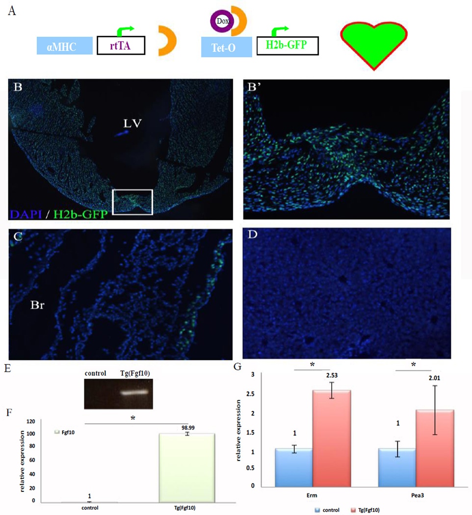
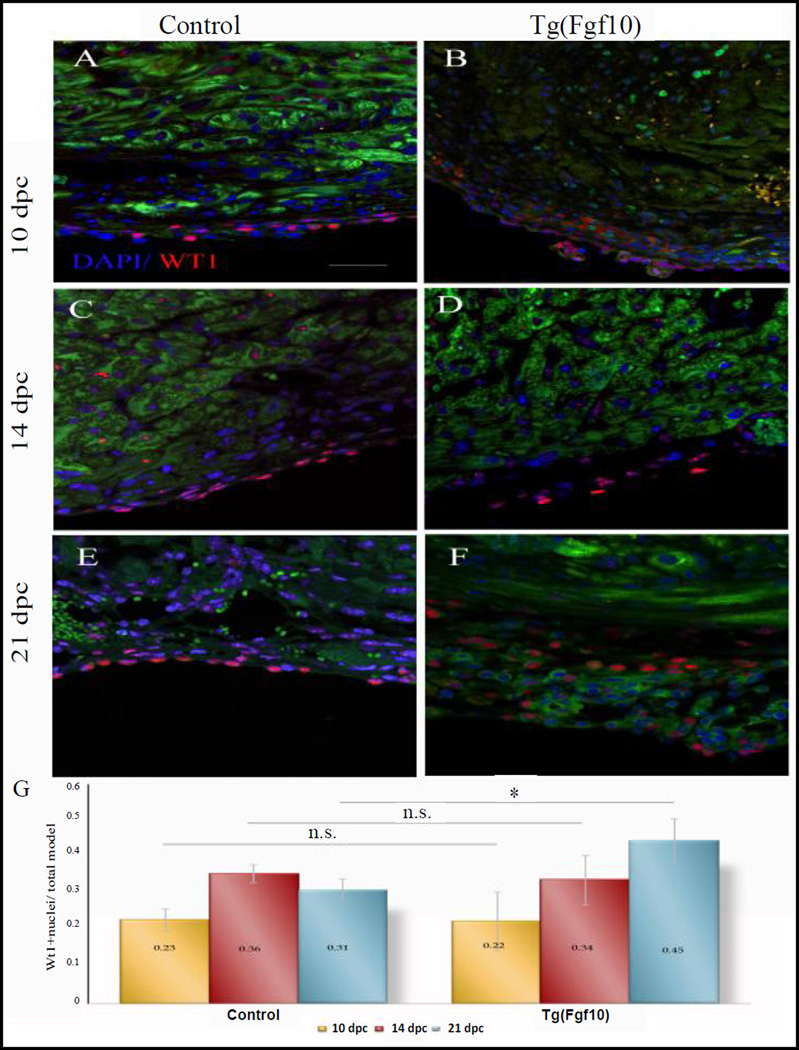
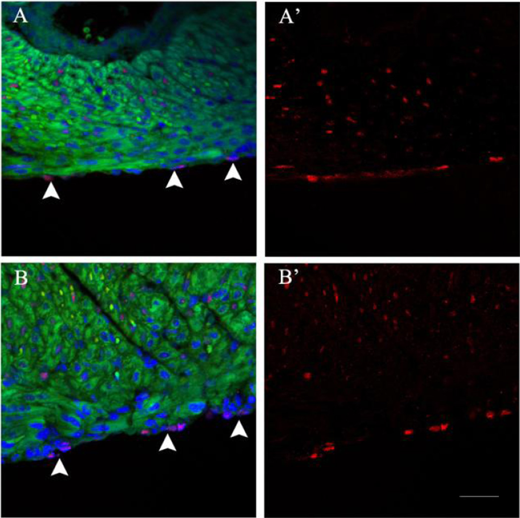
Unlike zebrafish and newt hearts, mammalian hearts have limited capacity to regenerate. Upon injury or disease, the adult mammalian hearts form a fibrotic scar. Recently, it was shown that neonatal mouse hearts can regenerate similarly to adult zebrafish hearts. However, this capacity quickly decreases after postnatal day 7 (P7). Understanding the molecular mechanisms underlying neonatal heart regeneration might lead to therapeutic approaches for regenerating adult mammalian hearts. In this study, we utilized an inducible transgenic mouse model to determine the effects of FGF10 growth factor over expression on neonatal mouse heart regeneration/repair. Over expression of FGF10 in myocardium enhanced the expansion of Wt1 positive epicardial cells at 21 days after heart injury through increased proliferation. However, this expansion of epicardial cells did not lead to increased epithelial-to-mesenchymal transition or affect fibroblast formation or fibrosis, as seen by vimentin expression, after heart injury. Furthermore, neither continuous nor transient expression of FGF10 did not affect scar thickness or length after heart injury in neonatal hearts. Our results suggest that FGF10 can regulate epicardial cell expansion of neonatal mouse hearts after injury; however, FGF10 alone is not sufficient to cause beneficial effects on heart repair.


 分享
分享

 求助内容:
求助内容: 应助结果提醒方式:
 扫码关注我们
