Gaetano Vattemi, Massimiliano Mirabella, Valeria Guglielmi, Matteo Lucchini, Giuliano Tomelleri, Anna Ghirardello, Andrea Doria
{"title":"特发性炎性肌病的肌肉活检特征及鉴别诊断。","authors":"Gaetano Vattemi, Massimiliano Mirabella, Valeria Guglielmi, Matteo Lucchini, Giuliano Tomelleri, Anna Ghirardello, Andrea Doria","doi":"10.1007/s13317-014-0062-2","DOIUrl":null,"url":null,"abstract":"<p><p>The gold standard to characterize idiopathic inflammatory myopathies is the morphological, immunohistochemical and immunopathological analysis of muscle biopsy. Mononuclear cell infiltrates and muscle fiber necrosis are commonly shared histopathological features. Inflammatory cells that surround, invade and destroy healthy muscle fibers expressing MHC class I antigen are the typical pathological finding of polymyositis. Perifascicular atrophy and microangiopathy strongly support a diagnosis of dermatomyositis. Randomly distributed necrotic muscle fibers without mononuclear cell infiltrates represent the histopathological hallmark of immune-mediated necrotizing myopathy; meanwhile, endomysial inflammation and muscle fiber degeneration are the two main pathological features in sporadic inclusion body myositis. A correct differential diagnosis requires immunopathological analysis of the muscle biopsy and has important clinical implications for therapeutic approach. In particular, unnecessary, potentially harmful, immune-suppressive therapy should be avoided alike in dystrophic myopathies with secondary inflammation. </p>","PeriodicalId":8655,"journal":{"name":"Auto-Immunity Highlights","volume":"5 3","pages":"77-85"},"PeriodicalIF":0.0000,"publicationDate":"2014-09-10","publicationTypes":"Journal Article","fieldsOfStudy":null,"isOpenAccess":false,"openAccessPdf":"https://sci-hub-pdf.com/10.1007/s13317-014-0062-2","citationCount":"70","resultStr":"{\"title\":\"Muscle biopsy features of idiopathic inflammatory myopathies and differential diagnosis.\",\"authors\":\"Gaetano Vattemi, Massimiliano Mirabella, Valeria Guglielmi, Matteo Lucchini, Giuliano Tomelleri, Anna Ghirardello, Andrea Doria\",\"doi\":\"10.1007/s13317-014-0062-2\",\"DOIUrl\":null,\"url\":null,\"abstract\":\"<p><p>The gold standard to characterize idiopathic inflammatory myopathies is the morphological, immunohistochemical and immunopathological analysis of muscle biopsy. Mononuclear cell infiltrates and muscle fiber necrosis are commonly shared histopathological features. Inflammatory cells that surround, invade and destroy healthy muscle fibers expressing MHC class I antigen are the typical pathological finding of polymyositis. Perifascicular atrophy and microangiopathy strongly support a diagnosis of dermatomyositis. Randomly distributed necrotic muscle fibers without mononuclear cell infiltrates represent the histopathological hallmark of immune-mediated necrotizing myopathy; meanwhile, endomysial inflammation and muscle fiber degeneration are the two main pathological features in sporadic inclusion body myositis. A correct differential diagnosis requires immunopathological analysis of the muscle biopsy and has important clinical implications for therapeutic approach. In particular, unnecessary, potentially harmful, immune-suppressive therapy should be avoided alike in dystrophic myopathies with secondary inflammation. </p>\",\"PeriodicalId\":8655,\"journal\":{\"name\":\"Auto-Immunity Highlights\",\"volume\":\"5 3\",\"pages\":\"77-85\"},\"PeriodicalIF\":0.0000,\"publicationDate\":\"2014-09-10\",\"publicationTypes\":\"Journal Article\",\"fieldsOfStudy\":null,\"isOpenAccess\":false,\"openAccessPdf\":\"https://sci-hub-pdf.com/10.1007/s13317-014-0062-2\",\"citationCount\":\"70\",\"resultStr\":null,\"platform\":\"Semanticscholar\",\"paperid\":null,\"PeriodicalName\":\"Auto-Immunity Highlights\",\"FirstCategoryId\":\"1085\",\"ListUrlMain\":\"https://doi.org/10.1007/s13317-014-0062-2\",\"RegionNum\":0,\"RegionCategory\":null,\"ArticlePicture\":[],\"TitleCN\":null,\"AbstractTextCN\":null,\"PMCID\":null,\"EPubDate\":\"2014/12/1 0:00:00\",\"PubModel\":\"eCollection\",\"JCR\":\"Q1\",\"JCRName\":\"Medicine\",\"Score\":null,\"Total\":0}","platform":"Semanticscholar","paperid":null,"PeriodicalName":"Auto-Immunity Highlights","FirstCategoryId":"1085","ListUrlMain":"https://doi.org/10.1007/s13317-014-0062-2","RegionNum":0,"RegionCategory":null,"ArticlePicture":[],"TitleCN":null,"AbstractTextCN":null,"PMCID":null,"EPubDate":"2014/12/1 0:00:00","PubModel":"eCollection","JCR":"Q1","JCRName":"Medicine","Score":null,"Total":0}
Muscle biopsy features of idiopathic inflammatory myopathies and differential diagnosis.
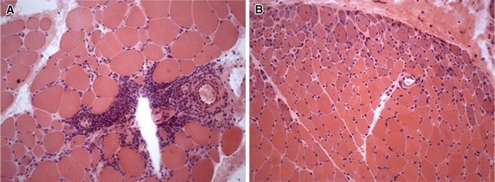
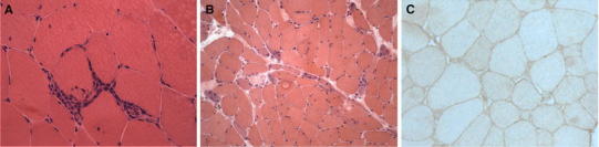
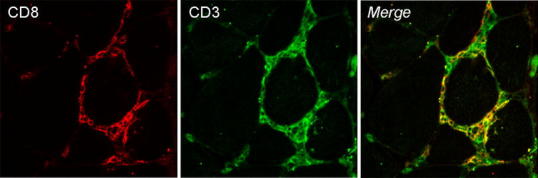
The gold standard to characterize idiopathic inflammatory myopathies is the morphological, immunohistochemical and immunopathological analysis of muscle biopsy. Mononuclear cell infiltrates and muscle fiber necrosis are commonly shared histopathological features. Inflammatory cells that surround, invade and destroy healthy muscle fibers expressing MHC class I antigen are the typical pathological finding of polymyositis. Perifascicular atrophy and microangiopathy strongly support a diagnosis of dermatomyositis. Randomly distributed necrotic muscle fibers without mononuclear cell infiltrates represent the histopathological hallmark of immune-mediated necrotizing myopathy; meanwhile, endomysial inflammation and muscle fiber degeneration are the two main pathological features in sporadic inclusion body myositis. A correct differential diagnosis requires immunopathological analysis of the muscle biopsy and has important clinical implications for therapeutic approach. In particular, unnecessary, potentially harmful, immune-suppressive therapy should be avoided alike in dystrophic myopathies with secondary inflammation.


 分享
分享
 求助内容:
求助内容: 应助结果提醒方式:
 扫码关注我们
