{"title":"特发性炎性肌炎的PET-MRI:临床和免疫标记物与影像学表现的比较研究。","authors":"Manu Santhappan Girija, Ravindu Tiwari, Seena Vengalil, Saraswati Nashi, Veeramani Preethish-Kumar, Kiran Polavarapu, Karthik Kulanthaivelu, Arpana Arbind, Mainak Bardhan, Akshata Huddar, Gopikrishnan Unnikrishnan, Valasani Ravi Kiran, Tanushree Chawla, Bevinahalli Nandeesh, Chandana Nagaraj, Atchayaram Nalini","doi":"10.1186/s42466-022-00213-9","DOIUrl":null,"url":null,"abstract":"<p><strong>Background: </strong>We sought to determine the utility of PET-MRI in diagnosing Idiopathic Inflammatory Myositis (IIM), and look for association between FDG uptake and clinical, pathological and laboratory parameters.</p><p><strong>Methods: </strong>A retrospective, observational study was conducted on IIM patients having positive serum autoantibodies and who underwent PET-MRI (3-Tesla SIEMENS Biograph MR scanner) between 2017 and 2021. Thirty patients who underwent PET-MRI to detect systemic metastasis without muscle involvement formed the control group.</p><p><strong>Results: </strong>In the IIM cohort, female: male sex ratio was 1.73, mean age at diagnosis was 40.33 years, and the mean duration of illness was 7 months. 33.33% of patients had severe limb weakness. Mi2B (43.33%), Mi2A (43.33%), PL-7(10%), PL-12(6.67%), SRP (16.67%), Tif1gamma (3.33%), NxP2 (3.33%), Ro-52(40%), PM-Scl, U1-RNP, ANA (26.67%) were the serum autoantibodies identified. Using SUV max Ratio to quantify FDG uptake, PET-MRI showed a sensitivity of 100% with 93.3% specificity in diagnosing IIM.FDG uptake was maximum in proximal lower limb region followed by proximal upper limb. Multivariate regression analysis showed that the severity of muscle weakness, serum Mi2B antibody positivity and serum creatinine kinase levels had a significant positive correlation with FDG uptake (value of 0.005, 0.043, 0.042, respectively for whole-body FDG uptake). FDG uptake also showed good correlation with histopathological features and muscle MRI, but there was no significant association with treatment response. Three female patients in our cohort had primary malignancy involving the breast, uterus, and cervix.</p><p><strong>Conclusions: </strong>PET-MRI is a promising diagnostic modality for IIM. PET-MRI reflects the severity of muscle inflammation, showing good association with various clinical/laboratory parameters, histopathology, and muscle MRI. Parameters associated with severe muscle inflammation in PET-MRI-clinical severity of muscle weakness, Mi2B positivity, and serum creatine kinase levels-may be used as clinical/laboratory markers of disease severity in IIM. PET-MRI has the added advantage of detection of systemic malignancy.</p>","PeriodicalId":19169,"journal":{"name":"Neurological Research and Practice","volume":" ","pages":"49"},"PeriodicalIF":0.0000,"publicationDate":"2022-10-10","publicationTypes":"Journal Article","fieldsOfStudy":null,"isOpenAccess":false,"openAccessPdf":"https://www.ncbi.nlm.nih.gov/pmc/articles/PMC9549636/pdf/","citationCount":"3","resultStr":"{\"title\":\"PET-MRI in idiopathic inflammatory myositis: a comparative study of clinical and immunological markers with imaging findings.\",\"authors\":\"Manu Santhappan Girija, Ravindu Tiwari, Seena Vengalil, Saraswati Nashi, Veeramani Preethish-Kumar, Kiran Polavarapu, Karthik Kulanthaivelu, Arpana Arbind, Mainak Bardhan, Akshata Huddar, Gopikrishnan Unnikrishnan, Valasani Ravi Kiran, Tanushree Chawla, Bevinahalli Nandeesh, Chandana Nagaraj, Atchayaram Nalini\",\"doi\":\"10.1186/s42466-022-00213-9\",\"DOIUrl\":null,\"url\":null,\"abstract\":\"<p><strong>Background: </strong>We sought to determine the utility of PET-MRI in diagnosing Idiopathic Inflammatory Myositis (IIM), and look for association between FDG uptake and clinical, pathological and laboratory parameters.</p><p><strong>Methods: </strong>A retrospective, observational study was conducted on IIM patients having positive serum autoantibodies and who underwent PET-MRI (3-Tesla SIEMENS Biograph MR scanner) between 2017 and 2021. Thirty patients who underwent PET-MRI to detect systemic metastasis without muscle involvement formed the control group.</p><p><strong>Results: </strong>In the IIM cohort, female: male sex ratio was 1.73, mean age at diagnosis was 40.33 years, and the mean duration of illness was 7 months. 33.33% of patients had severe limb weakness. Mi2B (43.33%), Mi2A (43.33%), PL-7(10%), PL-12(6.67%), SRP (16.67%), Tif1gamma (3.33%), NxP2 (3.33%), Ro-52(40%), PM-Scl, U1-RNP, ANA (26.67%) were the serum autoantibodies identified. Using SUV max Ratio to quantify FDG uptake, PET-MRI showed a sensitivity of 100% with 93.3% specificity in diagnosing IIM.FDG uptake was maximum in proximal lower limb region followed by proximal upper limb. Multivariate regression analysis showed that the severity of muscle weakness, serum Mi2B antibody positivity and serum creatinine kinase levels had a significant positive correlation with FDG uptake (value of 0.005, 0.043, 0.042, respectively for whole-body FDG uptake). FDG uptake also showed good correlation with histopathological features and muscle MRI, but there was no significant association with treatment response. Three female patients in our cohort had primary malignancy involving the breast, uterus, and cervix.</p><p><strong>Conclusions: </strong>PET-MRI is a promising diagnostic modality for IIM. PET-MRI reflects the severity of muscle inflammation, showing good association with various clinical/laboratory parameters, histopathology, and muscle MRI. Parameters associated with severe muscle inflammation in PET-MRI-clinical severity of muscle weakness, Mi2B positivity, and serum creatine kinase levels-may be used as clinical/laboratory markers of disease severity in IIM. PET-MRI has the added advantage of detection of systemic malignancy.</p>\",\"PeriodicalId\":19169,\"journal\":{\"name\":\"Neurological Research and Practice\",\"volume\":\" \",\"pages\":\"49\"},\"PeriodicalIF\":0.0000,\"publicationDate\":\"2022-10-10\",\"publicationTypes\":\"Journal Article\",\"fieldsOfStudy\":null,\"isOpenAccess\":false,\"openAccessPdf\":\"https://www.ncbi.nlm.nih.gov/pmc/articles/PMC9549636/pdf/\",\"citationCount\":\"3\",\"resultStr\":null,\"platform\":\"Semanticscholar\",\"paperid\":null,\"PeriodicalName\":\"Neurological Research and Practice\",\"FirstCategoryId\":\"1085\",\"ListUrlMain\":\"https://doi.org/10.1186/s42466-022-00213-9\",\"RegionNum\":0,\"RegionCategory\":null,\"ArticlePicture\":[],\"TitleCN\":null,\"AbstractTextCN\":null,\"PMCID\":null,\"EPubDate\":\"\",\"PubModel\":\"\",\"JCR\":\"\",\"JCRName\":\"\",\"Score\":null,\"Total\":0}","platform":"Semanticscholar","paperid":null,"PeriodicalName":"Neurological Research and Practice","FirstCategoryId":"1085","ListUrlMain":"https://doi.org/10.1186/s42466-022-00213-9","RegionNum":0,"RegionCategory":null,"ArticlePicture":[],"TitleCN":null,"AbstractTextCN":null,"PMCID":null,"EPubDate":"","PubModel":"","JCR":"","JCRName":"","Score":null,"Total":0}
引用次数: 3
摘要
背景:我们试图确定PET-MRI在诊断特发性炎症性肌炎(IIM)中的应用,并寻找FDG摄取与临床、病理和实验室参数之间的关系。方法:对2017年至2021年间接受PET-MRI (3-Tesla SIEMENS Biograph MR扫描仪)检查的血清自身抗体阳性的IIM患者进行回顾性观察研究。30例患者接受PET-MRI检查全身转移而不累及肌肉作为对照组。结果:IIM队列中男女性别比为1.73,平均诊断年龄为40.33岁,平均病程为7个月。33.33%的患者存在严重肢体无力。血清自身抗体分别为Mi2B(43.33%)、Mi2A(43.33%)、PL-7(10%)、PL-12(6.67%)、SRP(16.67%)、Tif1gamma(3.33%)、NxP2(3.33%)、Ro-52(40%)、PM-Scl、U1-RNP、ANA(26.67%)。利用SUV max Ratio量化FDG摄取,PET-MRI诊断IIM的敏感性为100%,特异性为93.3%。FDG摄取在下肢近端区域最大,其次是上肢近端。多因素回归分析显示,肌肉无力程度、血清Mi2B抗体阳性、血清肌酐激酶水平与FDG摄取呈显著正相关(全身FDG摄取值分别为0.005、0.043、0.042)。FDG摄取也与组织病理学特征和肌肉MRI表现出良好的相关性,但与治疗反应无显著相关性。在我们的队列中,有3名女性患者原发恶性肿瘤累及乳房、子宫和子宫颈。结论:PET-MRI是一种很有前途的IIM诊断方法。PET-MRI反映肌肉炎症的严重程度,与各种临床/实验室参数、组织病理学和肌肉MRI表现出良好的相关性。pet - mri中与严重肌肉炎症相关的参数(肌肉无力的临床严重程度、Mi2B阳性和血清肌酸激酶水平)可作为IIM疾病严重程度的临床/实验室标志物。PET-MRI具有检测全身恶性肿瘤的附加优势。
PET-MRI in idiopathic inflammatory myositis: a comparative study of clinical and immunological markers with imaging findings.
Background: We sought to determine the utility of PET-MRI in diagnosing Idiopathic Inflammatory Myositis (IIM), and look for association between FDG uptake and clinical, pathological and laboratory parameters.
Methods: A retrospective, observational study was conducted on IIM patients having positive serum autoantibodies and who underwent PET-MRI (3-Tesla SIEMENS Biograph MR scanner) between 2017 and 2021. Thirty patients who underwent PET-MRI to detect systemic metastasis without muscle involvement formed the control group.
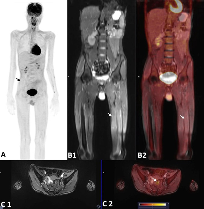
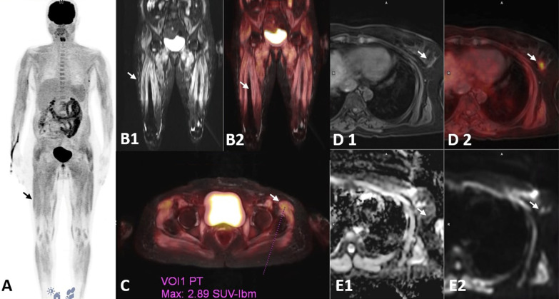
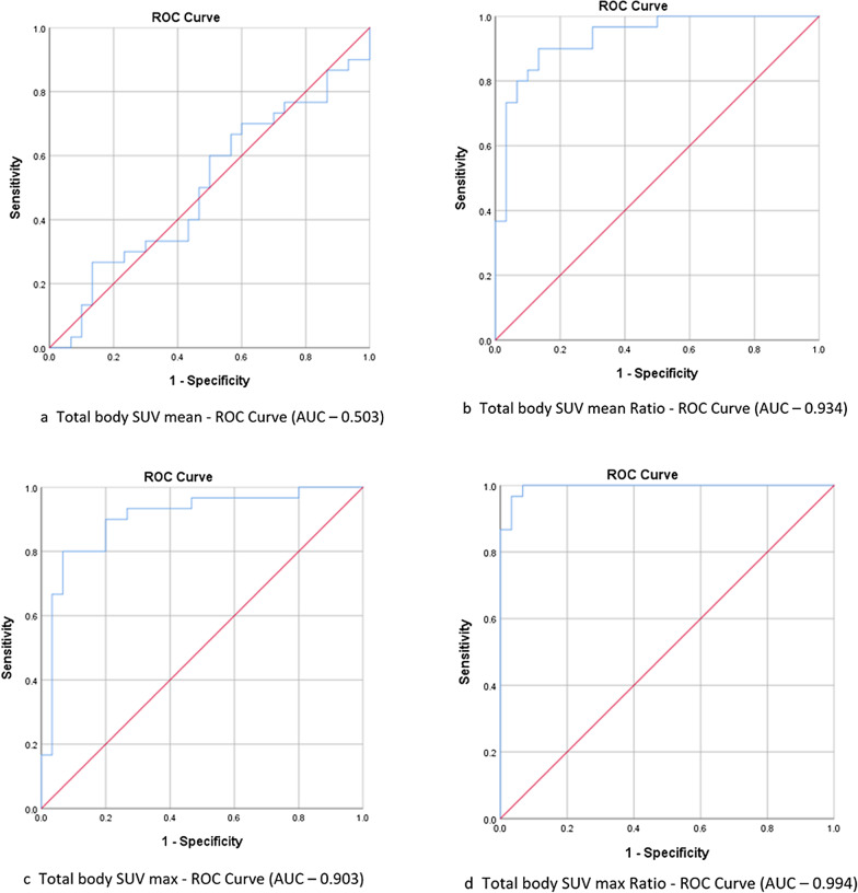
Results: In the IIM cohort, female: male sex ratio was 1.73, mean age at diagnosis was 40.33 years, and the mean duration of illness was 7 months. 33.33% of patients had severe limb weakness. Mi2B (43.33%), Mi2A (43.33%), PL-7(10%), PL-12(6.67%), SRP (16.67%), Tif1gamma (3.33%), NxP2 (3.33%), Ro-52(40%), PM-Scl, U1-RNP, ANA (26.67%) were the serum autoantibodies identified. Using SUV max Ratio to quantify FDG uptake, PET-MRI showed a sensitivity of 100% with 93.3% specificity in diagnosing IIM.FDG uptake was maximum in proximal lower limb region followed by proximal upper limb. Multivariate regression analysis showed that the severity of muscle weakness, serum Mi2B antibody positivity and serum creatinine kinase levels had a significant positive correlation with FDG uptake (value of 0.005, 0.043, 0.042, respectively for whole-body FDG uptake). FDG uptake also showed good correlation with histopathological features and muscle MRI, but there was no significant association with treatment response. Three female patients in our cohort had primary malignancy involving the breast, uterus, and cervix.
Conclusions: PET-MRI is a promising diagnostic modality for IIM. PET-MRI reflects the severity of muscle inflammation, showing good association with various clinical/laboratory parameters, histopathology, and muscle MRI. Parameters associated with severe muscle inflammation in PET-MRI-clinical severity of muscle weakness, Mi2B positivity, and serum creatine kinase levels-may be used as clinical/laboratory markers of disease severity in IIM. PET-MRI has the added advantage of detection of systemic malignancy.


 分享
分享

 求助内容:
求助内容: 应助结果提醒方式:
 扫码关注我们
