Yeo Eun Han, Beom Jin Park, Deuk Jae Sung, Min Ju Kim, Na Yeon Han, Ki Choon Sim, Yongwon Cho, Hayeon Kim
{"title":"胰腺导管腺癌的双层光谱CT:门静脉期的虚拟单能图像可以替代胰期扫描吗?","authors":"Yeo Eun Han, Beom Jin Park, Deuk Jae Sung, Min Ju Kim, Na Yeon Han, Ki Choon Sim, Yongwon Cho, Hayeon Kim","doi":"10.5334/jbsr.2798","DOIUrl":null,"url":null,"abstract":"<p><strong>Objectives: </strong>To determine the performance of virtual monoenergetic images (VMIs) of the portal venous phase (PVP) compared with the pancreatic-phase image for pancreatic ductal adenocarcinoma (PDAC) evaluation.</p><p><strong>Materials and methods: </strong>This retrospective study enrolled 64 patients with PDAC who underwent pancreatic CT with dual-layer spectral CT between February 2018 and January 2020. A polychromatic pancreatic-phase image and VMIs at 40 (VMI<sub>40</sub>), 55 (VMI<sub>55</sub>), and 70 keV (VMI<sub>70</sub>) of the PVP were generated. The tumor-to-pancreas contrast-to-noise ratio (CNR), attenuation difference, peripancreatic vascular signal-to-noise ratio (SNR), and CNR were compared among the four images. Subjective image analysis was performed for tumor conspicuity, heterogeneity, size, and arterial invasion.</p><p><strong>Results: </strong>VMI<sub>40</sub> and VMI<sub>55</sub> demonstrated higher tumor-to-pancreas CNR, attenuation difference, and higher peripancreatic vascular CNR and SNR than the pancreatic-phase image and VMI<sub>70</sub> (p < .001). On subjective analysis, VMI<sub>55</sub> showed the best tumor conspicuity. Moreover, the inter-reader agreement for arterial invasion in VMIs from the PVP was not inferior to that in the pancreatic-phase image.</p><p><strong>Conclusion: </strong>For evaluating PDAC, the VMI<sub>55</sub> of the PVP was superior to the pancreatic-phase image in terms of tumor conspicuity and peripancreatic vascular enhancement. Therefore, the VMI<sub>55</sub> of the PVP could be an alternative to the pancreatic-phase scan in patients suspicious of PDAC.</p>","PeriodicalId":56282,"journal":{"name":"Journal of the Belgian Society of Radiology","volume":"106 1","pages":"83"},"PeriodicalIF":1.3000,"publicationDate":"2022-09-22","publicationTypes":"Journal Article","fieldsOfStudy":null,"isOpenAccess":false,"openAccessPdf":"https://www.ncbi.nlm.nih.gov/pmc/articles/PMC9504095/pdf/","citationCount":"3","resultStr":"{\"title\":\"Dual-Layer Spectral CT of Pancreas Ductal Adenocarcinoma: Can Virtual Monoenergetic Images of the Portal Venous Phase Be an Alternative to the Pancreatic-Phase Scan?\",\"authors\":\"Yeo Eun Han, Beom Jin Park, Deuk Jae Sung, Min Ju Kim, Na Yeon Han, Ki Choon Sim, Yongwon Cho, Hayeon Kim\",\"doi\":\"10.5334/jbsr.2798\",\"DOIUrl\":null,\"url\":null,\"abstract\":\"<p><strong>Objectives: </strong>To determine the performance of virtual monoenergetic images (VMIs) of the portal venous phase (PVP) compared with the pancreatic-phase image for pancreatic ductal adenocarcinoma (PDAC) evaluation.</p><p><strong>Materials and methods: </strong>This retrospective study enrolled 64 patients with PDAC who underwent pancreatic CT with dual-layer spectral CT between February 2018 and January 2020. A polychromatic pancreatic-phase image and VMIs at 40 (VMI<sub>40</sub>), 55 (VMI<sub>55</sub>), and 70 keV (VMI<sub>70</sub>) of the PVP were generated. The tumor-to-pancreas contrast-to-noise ratio (CNR), attenuation difference, peripancreatic vascular signal-to-noise ratio (SNR), and CNR were compared among the four images. Subjective image analysis was performed for tumor conspicuity, heterogeneity, size, and arterial invasion.</p><p><strong>Results: </strong>VMI<sub>40</sub> and VMI<sub>55</sub> demonstrated higher tumor-to-pancreas CNR, attenuation difference, and higher peripancreatic vascular CNR and SNR than the pancreatic-phase image and VMI<sub>70</sub> (p < .001). On subjective analysis, VMI<sub>55</sub> showed the best tumor conspicuity. Moreover, the inter-reader agreement for arterial invasion in VMIs from the PVP was not inferior to that in the pancreatic-phase image.</p><p><strong>Conclusion: </strong>For evaluating PDAC, the VMI<sub>55</sub> of the PVP was superior to the pancreatic-phase image in terms of tumor conspicuity and peripancreatic vascular enhancement. Therefore, the VMI<sub>55</sub> of the PVP could be an alternative to the pancreatic-phase scan in patients suspicious of PDAC.</p>\",\"PeriodicalId\":56282,\"journal\":{\"name\":\"Journal of the Belgian Society of Radiology\",\"volume\":\"106 1\",\"pages\":\"83\"},\"PeriodicalIF\":1.3000,\"publicationDate\":\"2022-09-22\",\"publicationTypes\":\"Journal Article\",\"fieldsOfStudy\":null,\"isOpenAccess\":false,\"openAccessPdf\":\"https://www.ncbi.nlm.nih.gov/pmc/articles/PMC9504095/pdf/\",\"citationCount\":\"3\",\"resultStr\":null,\"platform\":\"Semanticscholar\",\"paperid\":null,\"PeriodicalName\":\"Journal of the Belgian Society of Radiology\",\"FirstCategoryId\":\"3\",\"ListUrlMain\":\"https://doi.org/10.5334/jbsr.2798\",\"RegionNum\":4,\"RegionCategory\":\"医学\",\"ArticlePicture\":[],\"TitleCN\":null,\"AbstractTextCN\":null,\"PMCID\":null,\"EPubDate\":\"2022/1/1 0:00:00\",\"PubModel\":\"eCollection\",\"JCR\":\"Q4\",\"JCRName\":\"Medicine\",\"Score\":null,\"Total\":0}","platform":"Semanticscholar","paperid":null,"PeriodicalName":"Journal of the Belgian Society of Radiology","FirstCategoryId":"3","ListUrlMain":"https://doi.org/10.5334/jbsr.2798","RegionNum":4,"RegionCategory":"医学","ArticlePicture":[],"TitleCN":null,"AbstractTextCN":null,"PMCID":null,"EPubDate":"2022/1/1 0:00:00","PubModel":"eCollection","JCR":"Q4","JCRName":"Medicine","Score":null,"Total":0}
Dual-Layer Spectral CT of Pancreas Ductal Adenocarcinoma: Can Virtual Monoenergetic Images of the Portal Venous Phase Be an Alternative to the Pancreatic-Phase Scan?
Objectives: To determine the performance of virtual monoenergetic images (VMIs) of the portal venous phase (PVP) compared with the pancreatic-phase image for pancreatic ductal adenocarcinoma (PDAC) evaluation.
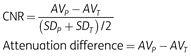
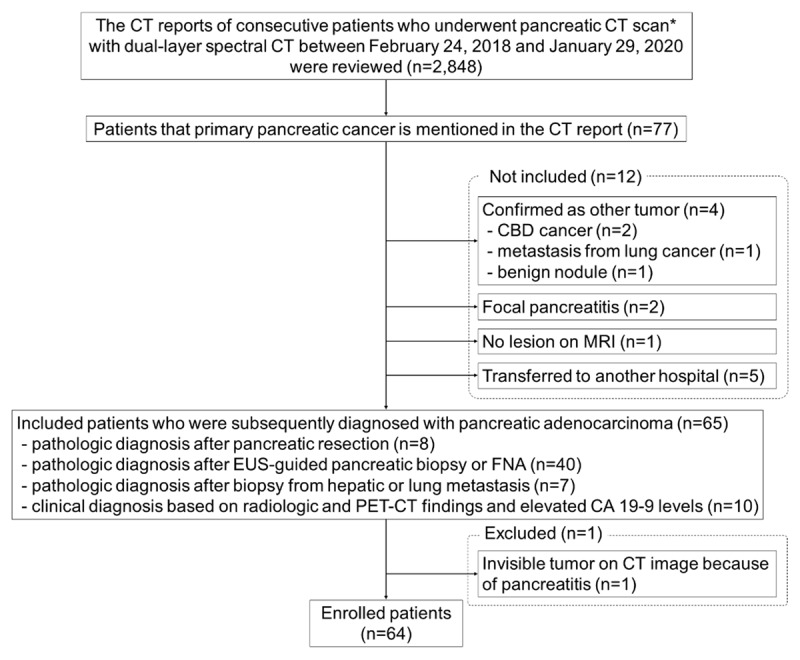
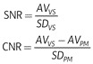
Materials and methods: This retrospective study enrolled 64 patients with PDAC who underwent pancreatic CT with dual-layer spectral CT between February 2018 and January 2020. A polychromatic pancreatic-phase image and VMIs at 40 (VMI40), 55 (VMI55), and 70 keV (VMI70) of the PVP were generated. The tumor-to-pancreas contrast-to-noise ratio (CNR), attenuation difference, peripancreatic vascular signal-to-noise ratio (SNR), and CNR were compared among the four images. Subjective image analysis was performed for tumor conspicuity, heterogeneity, size, and arterial invasion.
Results: VMI40 and VMI55 demonstrated higher tumor-to-pancreas CNR, attenuation difference, and higher peripancreatic vascular CNR and SNR than the pancreatic-phase image and VMI70 (p < .001). On subjective analysis, VMI55 showed the best tumor conspicuity. Moreover, the inter-reader agreement for arterial invasion in VMIs from the PVP was not inferior to that in the pancreatic-phase image.
Conclusion: For evaluating PDAC, the VMI55 of the PVP was superior to the pancreatic-phase image in terms of tumor conspicuity and peripancreatic vascular enhancement. Therefore, the VMI55 of the PVP could be an alternative to the pancreatic-phase scan in patients suspicious of PDAC.
期刊介绍:
The purpose of the Journal of the Belgian Society of Radiology is the publication of articles dealing with diagnostic and interventional radiology, related imaging techniques, allied sciences, and continuing education.


 分享
分享
 求助内容:
求助内容: 应助结果提醒方式:
 扫码关注我们
