Hee Soo Won, Yoon Ki Cha, Jeung Sook Kim, Seo Jin Jang, So Hyeon Bak, Hyun Jung Yoon
{"title":"胸腔异物的影像学表现综述。","authors":"Hee Soo Won, Yoon Ki Cha, Jeung Sook Kim, Seo Jin Jang, So Hyeon Bak, Hyun Jung Yoon","doi":"10.3348/jksr.2021.0084","DOIUrl":null,"url":null,"abstract":"<p><p>Thoracic foreign bodies (FBs) are serious and relatively frequent in emergency departments. Thoracic FBs may occur in association with aspiration, ingestion, trauma, or iatrogenic causes. Imaging plays an important role in the identification of FBs and their dimensions, structures, and locations, before the initiation of interventional treatment. To guide proper clinical management, radiologists should be aware of the radiologic presentations and the consequences of thoracic FBs. In this pictorial essay, we reviewed the optimal imaging settings to identify FBs in the thorax, classified thoracic FBs into four types according to their etiology, and reviewed the characteristic imaging features and the possible complications.</p>","PeriodicalId":74904,"journal":{"name":"Taehan Yongsang Uihakhoe chi","volume":" ","pages":"293-303"},"PeriodicalIF":0.0000,"publicationDate":"2022-03-01","publicationTypes":"Journal Article","fieldsOfStudy":null,"isOpenAccess":false,"openAccessPdf":"https://ftp.ncbi.nlm.nih.gov/pub/pmc/oa_pdf/c3/1f/jksr-83-293.PMC9514446.pdf","citationCount":"0","resultStr":"{\"title\":\"A Pictorial Review of Radiologic Findings of Foreign Bodies in the Thorax.\",\"authors\":\"Hee Soo Won, Yoon Ki Cha, Jeung Sook Kim, Seo Jin Jang, So Hyeon Bak, Hyun Jung Yoon\",\"doi\":\"10.3348/jksr.2021.0084\",\"DOIUrl\":null,\"url\":null,\"abstract\":\"<p><p>Thoracic foreign bodies (FBs) are serious and relatively frequent in emergency departments. Thoracic FBs may occur in association with aspiration, ingestion, trauma, or iatrogenic causes. Imaging plays an important role in the identification of FBs and their dimensions, structures, and locations, before the initiation of interventional treatment. To guide proper clinical management, radiologists should be aware of the radiologic presentations and the consequences of thoracic FBs. In this pictorial essay, we reviewed the optimal imaging settings to identify FBs in the thorax, classified thoracic FBs into four types according to their etiology, and reviewed the characteristic imaging features and the possible complications.</p>\",\"PeriodicalId\":74904,\"journal\":{\"name\":\"Taehan Yongsang Uihakhoe chi\",\"volume\":\" \",\"pages\":\"293-303\"},\"PeriodicalIF\":0.0000,\"publicationDate\":\"2022-03-01\",\"publicationTypes\":\"Journal Article\",\"fieldsOfStudy\":null,\"isOpenAccess\":false,\"openAccessPdf\":\"https://ftp.ncbi.nlm.nih.gov/pub/pmc/oa_pdf/c3/1f/jksr-83-293.PMC9514446.pdf\",\"citationCount\":\"0\",\"resultStr\":null,\"platform\":\"Semanticscholar\",\"paperid\":null,\"PeriodicalName\":\"Taehan Yongsang Uihakhoe chi\",\"FirstCategoryId\":\"1085\",\"ListUrlMain\":\"https://doi.org/10.3348/jksr.2021.0084\",\"RegionNum\":0,\"RegionCategory\":null,\"ArticlePicture\":[],\"TitleCN\":null,\"AbstractTextCN\":null,\"PMCID\":null,\"EPubDate\":\"2021/9/27 0:00:00\",\"PubModel\":\"Epub\",\"JCR\":\"\",\"JCRName\":\"\",\"Score\":null,\"Total\":0}","platform":"Semanticscholar","paperid":null,"PeriodicalName":"Taehan Yongsang Uihakhoe chi","FirstCategoryId":"1085","ListUrlMain":"https://doi.org/10.3348/jksr.2021.0084","RegionNum":0,"RegionCategory":null,"ArticlePicture":[],"TitleCN":null,"AbstractTextCN":null,"PMCID":null,"EPubDate":"2021/9/27 0:00:00","PubModel":"Epub","JCR":"","JCRName":"","Score":null,"Total":0}
A Pictorial Review of Radiologic Findings of Foreign Bodies in the Thorax.
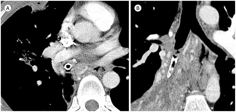
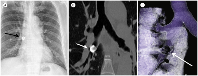
Thoracic foreign bodies (FBs) are serious and relatively frequent in emergency departments. Thoracic FBs may occur in association with aspiration, ingestion, trauma, or iatrogenic causes. Imaging plays an important role in the identification of FBs and their dimensions, structures, and locations, before the initiation of interventional treatment. To guide proper clinical management, radiologists should be aware of the radiologic presentations and the consequences of thoracic FBs. In this pictorial essay, we reviewed the optimal imaging settings to identify FBs in the thorax, classified thoracic FBs into four types according to their etiology, and reviewed the characteristic imaging features and the possible complications.


 分享
分享

 求助内容:
求助内容: 应助结果提醒方式:
 扫码关注我们
