Ana Gabriella de Oliveira Sardinha, Ceres Nunes de Resende Oyama, Armando de Mendonça Maroja, Ivan F Costa
{"title":"一种快速模拟纤维和流体形成的生物组织的变形方法的实现和临床应用。","authors":"Ana Gabriella de Oliveira Sardinha, Ceres Nunes de Resende Oyama, Armando de Mendonça Maroja, Ivan F Costa","doi":"10.1186/s13029-016-0054-x","DOIUrl":null,"url":null,"abstract":"<p><strong>Background: </strong>The aim of this paper is to provide a general discussion, algorithm, and actual working programs of the deformation method for fast simulation of biological tissue formed by fibers and fluid. In order to demonstrate the benefit of the clinical applications software, we successfully used our computational program to deform a 3D breast image acquired from patients, using a 3D scanner, in a real hospital environment.</p><p><strong>Results: </strong>The method implements a quasi-static solution for elastic global deformations of objects. Each pair of vertices of the surface is connected and defines an elastic fiber. The set of all the elastic fibers defines a mesh of smaller size than the volumetric meshes, allowing for simulation of complex objects with less computational effort. The behavior similar to the stress tensor is obtained by the volume conservation equation that mixes the 3D coordinates. Step by step, we show the computational implementation of this approach.</p><p><strong>Conclusions: </strong>As an example, a 2D rectangle formed by only 4 vertices is solved and, for this simple geometry, all intermediate results are shown. On the other hand, actual implementations of these ideas in the form of working computer routines are provided for general 3D objects, including a clinical application.</p>","PeriodicalId":35052,"journal":{"name":"Source Code for Biology and Medicine","volume":" ","pages":"7"},"PeriodicalIF":0.0000,"publicationDate":"2016-04-15","publicationTypes":"Journal Article","fieldsOfStudy":null,"isOpenAccess":false,"openAccessPdf":"https://sci-hub-pdf.com/10.1186/s13029-016-0054-x","citationCount":"0","resultStr":"{\"title\":\"Implementation and clinical application of a deformation method for fast simulation of biological tissue formed by fibers and fluid.\",\"authors\":\"Ana Gabriella de Oliveira Sardinha, Ceres Nunes de Resende Oyama, Armando de Mendonça Maroja, Ivan F Costa\",\"doi\":\"10.1186/s13029-016-0054-x\",\"DOIUrl\":null,\"url\":null,\"abstract\":\"<p><strong>Background: </strong>The aim of this paper is to provide a general discussion, algorithm, and actual working programs of the deformation method for fast simulation of biological tissue formed by fibers and fluid. In order to demonstrate the benefit of the clinical applications software, we successfully used our computational program to deform a 3D breast image acquired from patients, using a 3D scanner, in a real hospital environment.</p><p><strong>Results: </strong>The method implements a quasi-static solution for elastic global deformations of objects. Each pair of vertices of the surface is connected and defines an elastic fiber. The set of all the elastic fibers defines a mesh of smaller size than the volumetric meshes, allowing for simulation of complex objects with less computational effort. The behavior similar to the stress tensor is obtained by the volume conservation equation that mixes the 3D coordinates. Step by step, we show the computational implementation of this approach.</p><p><strong>Conclusions: </strong>As an example, a 2D rectangle formed by only 4 vertices is solved and, for this simple geometry, all intermediate results are shown. On the other hand, actual implementations of these ideas in the form of working computer routines are provided for general 3D objects, including a clinical application.</p>\",\"PeriodicalId\":35052,\"journal\":{\"name\":\"Source Code for Biology and Medicine\",\"volume\":\" \",\"pages\":\"7\"},\"PeriodicalIF\":0.0000,\"publicationDate\":\"2016-04-15\",\"publicationTypes\":\"Journal Article\",\"fieldsOfStudy\":null,\"isOpenAccess\":false,\"openAccessPdf\":\"https://sci-hub-pdf.com/10.1186/s13029-016-0054-x\",\"citationCount\":\"0\",\"resultStr\":null,\"platform\":\"Semanticscholar\",\"paperid\":null,\"PeriodicalName\":\"Source Code for Biology and Medicine\",\"FirstCategoryId\":\"1085\",\"ListUrlMain\":\"https://doi.org/10.1186/s13029-016-0054-x\",\"RegionNum\":0,\"RegionCategory\":null,\"ArticlePicture\":[],\"TitleCN\":null,\"AbstractTextCN\":null,\"PMCID\":null,\"EPubDate\":\"2016/1/1 0:00:00\",\"PubModel\":\"eCollection\",\"JCR\":\"Q2\",\"JCRName\":\"Decision Sciences\",\"Score\":null,\"Total\":0}","platform":"Semanticscholar","paperid":null,"PeriodicalName":"Source Code for Biology and Medicine","FirstCategoryId":"1085","ListUrlMain":"https://doi.org/10.1186/s13029-016-0054-x","RegionNum":0,"RegionCategory":null,"ArticlePicture":[],"TitleCN":null,"AbstractTextCN":null,"PMCID":null,"EPubDate":"2016/1/1 0:00:00","PubModel":"eCollection","JCR":"Q2","JCRName":"Decision Sciences","Score":null,"Total":0}
Implementation and clinical application of a deformation method for fast simulation of biological tissue formed by fibers and fluid.
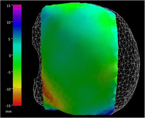
Background: The aim of this paper is to provide a general discussion, algorithm, and actual working programs of the deformation method for fast simulation of biological tissue formed by fibers and fluid. In order to demonstrate the benefit of the clinical applications software, we successfully used our computational program to deform a 3D breast image acquired from patients, using a 3D scanner, in a real hospital environment.
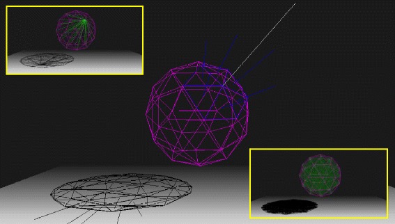
Results: The method implements a quasi-static solution for elastic global deformations of objects. Each pair of vertices of the surface is connected and defines an elastic fiber. The set of all the elastic fibers defines a mesh of smaller size than the volumetric meshes, allowing for simulation of complex objects with less computational effort. The behavior similar to the stress tensor is obtained by the volume conservation equation that mixes the 3D coordinates. Step by step, we show the computational implementation of this approach.
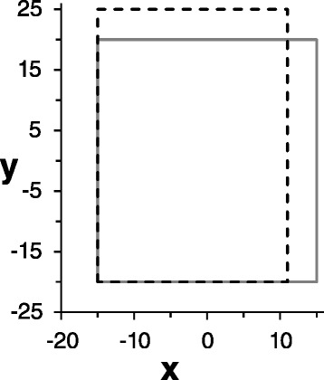
Conclusions: As an example, a 2D rectangle formed by only 4 vertices is solved and, for this simple geometry, all intermediate results are shown. On the other hand, actual implementations of these ideas in the form of working computer routines are provided for general 3D objects, including a clinical application.
期刊介绍:
Source Code for Biology and Medicine is a peer-reviewed open access, online journal that publishes articles on source code employed over a wide range of applications in biology and medicine. The journal"s aim is to publish source code for distribution and use in the public domain in order to advance biological and medical research. Through this dissemination, it may be possible to shorten the time required for solving certain computational problems for which there is limited source code availability or resources.


 分享
分享
 求助内容:
求助内容: 应助结果提醒方式:
 扫码关注我们
