Agazi G Gebreselassie, Delamo I Bekele, Yonette Paul, Julius S Ngwa, Daniel A Larbi
{"title":"评价晕厥在一个主要的黑人人口:重点神经影像学。","authors":"Agazi G Gebreselassie, Delamo I Bekele, Yonette Paul, Julius S Ngwa, Daniel A Larbi","doi":"10.4103/1947-2714.187133","DOIUrl":null,"url":null,"abstract":"<p><strong>Background: </strong>Current guidelines do not support the routine use of computed tomography (CT) scan of the head in the diagnostic workup of syncope. There is a lack of research to support whether these guidelines apply to the Black population.</p><p><strong>Aims: </strong>This study aims to evaluate the yield of neuroimaging in the evaluation of Syncope in a predominantly Black patient population and to test whether current guidelines based on studies conducted in other populations hold true in this group.</p><p><strong>Material and methods: </strong>A retrospective review of records of 151 patients admitted to a University Hospital with Syncope from 2011 to 2014 was performed. Data collected include CT head, magnetic resonance imaging of the brain, magnetic resonance angiogram, electroencephalogram, and orthostatic vital signs. Demographic data, admitting service, and comorbid conditions were identified. Syncope was classified as cardiogenic, orthostatic, vasovagal, situational, or undetermined. Statistical analysis was performed to determine which diagnostic tools were useful in identifying the potential causes of syncope. Data analysis was conducted using the Statistical Analysis System software 9.3 (SAS Institute, Cary, NC) and Statistical Analysis and Graphics (NCSS 9.0.7, Kaysville, UT).</p><p><strong>Results: </strong>One hundred and twenty eight (84.8%) of the patients were Black. The average age was 56.62 ± 18.78 standard deviation and 68.2% (103) were female. One hundred and fourteen patients (75.5%) had a CT brain. Five out of 114 patients had an acute abnormality on CT (4.4%). Only 1 of these 5 patients had an abnormality that was related to syncope. CT brain (P = 0.978) was not found to be predictive of underlying etiology of syncope despite high frequency of use.</p><p><strong>Conclusions: </strong>CT head was not useful in determining the etiology of syncope in a predominantly Black population. Current guidelines and studies conducted in other populations have detected similar findings.</p>","PeriodicalId":19703,"journal":{"name":"North American Journal of Medical Sciences","volume":"8 7","pages":"279-83"},"PeriodicalIF":0.0000,"publicationDate":"2016-07-01","publicationTypes":"Journal Article","fieldsOfStudy":null,"isOpenAccess":false,"openAccessPdf":"https://ftp.ncbi.nlm.nih.gov/pub/pmc/oa_pdf/81/6b/NAJMS-8-279.PMC4982356.pdf","citationCount":"2","resultStr":"{\"title\":\"The Evaluation of Syncope in a Predominantly Black Population: Focus on Neuroimaging.\",\"authors\":\"Agazi G Gebreselassie, Delamo I Bekele, Yonette Paul, Julius S Ngwa, Daniel A Larbi\",\"doi\":\"10.4103/1947-2714.187133\",\"DOIUrl\":null,\"url\":null,\"abstract\":\"<p><strong>Background: </strong>Current guidelines do not support the routine use of computed tomography (CT) scan of the head in the diagnostic workup of syncope. There is a lack of research to support whether these guidelines apply to the Black population.</p><p><strong>Aims: </strong>This study aims to evaluate the yield of neuroimaging in the evaluation of Syncope in a predominantly Black patient population and to test whether current guidelines based on studies conducted in other populations hold true in this group.</p><p><strong>Material and methods: </strong>A retrospective review of records of 151 patients admitted to a University Hospital with Syncope from 2011 to 2014 was performed. Data collected include CT head, magnetic resonance imaging of the brain, magnetic resonance angiogram, electroencephalogram, and orthostatic vital signs. Demographic data, admitting service, and comorbid conditions were identified. Syncope was classified as cardiogenic, orthostatic, vasovagal, situational, or undetermined. Statistical analysis was performed to determine which diagnostic tools were useful in identifying the potential causes of syncope. Data analysis was conducted using the Statistical Analysis System software 9.3 (SAS Institute, Cary, NC) and Statistical Analysis and Graphics (NCSS 9.0.7, Kaysville, UT).</p><p><strong>Results: </strong>One hundred and twenty eight (84.8%) of the patients were Black. The average age was 56.62 ± 18.78 standard deviation and 68.2% (103) were female. One hundred and fourteen patients (75.5%) had a CT brain. Five out of 114 patients had an acute abnormality on CT (4.4%). Only 1 of these 5 patients had an abnormality that was related to syncope. CT brain (P = 0.978) was not found to be predictive of underlying etiology of syncope despite high frequency of use.</p><p><strong>Conclusions: </strong>CT head was not useful in determining the etiology of syncope in a predominantly Black population. Current guidelines and studies conducted in other populations have detected similar findings.</p>\",\"PeriodicalId\":19703,\"journal\":{\"name\":\"North American Journal of Medical Sciences\",\"volume\":\"8 7\",\"pages\":\"279-83\"},\"PeriodicalIF\":0.0000,\"publicationDate\":\"2016-07-01\",\"publicationTypes\":\"Journal Article\",\"fieldsOfStudy\":null,\"isOpenAccess\":false,\"openAccessPdf\":\"https://ftp.ncbi.nlm.nih.gov/pub/pmc/oa_pdf/81/6b/NAJMS-8-279.PMC4982356.pdf\",\"citationCount\":\"2\",\"resultStr\":null,\"platform\":\"Semanticscholar\",\"paperid\":null,\"PeriodicalName\":\"North American Journal of Medical Sciences\",\"FirstCategoryId\":\"1085\",\"ListUrlMain\":\"https://doi.org/10.4103/1947-2714.187133\",\"RegionNum\":0,\"RegionCategory\":null,\"ArticlePicture\":[],\"TitleCN\":null,\"AbstractTextCN\":null,\"PMCID\":null,\"EPubDate\":\"\",\"PubModel\":\"\",\"JCR\":\"\",\"JCRName\":\"\",\"Score\":null,\"Total\":0}","platform":"Semanticscholar","paperid":null,"PeriodicalName":"North American Journal of Medical Sciences","FirstCategoryId":"1085","ListUrlMain":"https://doi.org/10.4103/1947-2714.187133","RegionNum":0,"RegionCategory":null,"ArticlePicture":[],"TitleCN":null,"AbstractTextCN":null,"PMCID":null,"EPubDate":"","PubModel":"","JCR":"","JCRName":"","Score":null,"Total":0}
The Evaluation of Syncope in a Predominantly Black Population: Focus on Neuroimaging.
Background: Current guidelines do not support the routine use of computed tomography (CT) scan of the head in the diagnostic workup of syncope. There is a lack of research to support whether these guidelines apply to the Black population.
Aims: This study aims to evaluate the yield of neuroimaging in the evaluation of Syncope in a predominantly Black patient population and to test whether current guidelines based on studies conducted in other populations hold true in this group.
Material and methods: A retrospective review of records of 151 patients admitted to a University Hospital with Syncope from 2011 to 2014 was performed. Data collected include CT head, magnetic resonance imaging of the brain, magnetic resonance angiogram, electroencephalogram, and orthostatic vital signs. Demographic data, admitting service, and comorbid conditions were identified. Syncope was classified as cardiogenic, orthostatic, vasovagal, situational, or undetermined. Statistical analysis was performed to determine which diagnostic tools were useful in identifying the potential causes of syncope. Data analysis was conducted using the Statistical Analysis System software 9.3 (SAS Institute, Cary, NC) and Statistical Analysis and Graphics (NCSS 9.0.7, Kaysville, UT).
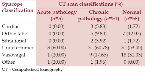
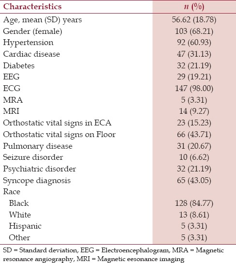
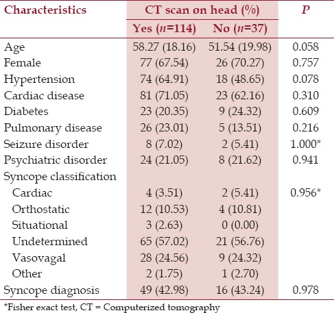
Results: One hundred and twenty eight (84.8%) of the patients were Black. The average age was 56.62 ± 18.78 standard deviation and 68.2% (103) were female. One hundred and fourteen patients (75.5%) had a CT brain. Five out of 114 patients had an acute abnormality on CT (4.4%). Only 1 of these 5 patients had an abnormality that was related to syncope. CT brain (P = 0.978) was not found to be predictive of underlying etiology of syncope despite high frequency of use.
Conclusions: CT head was not useful in determining the etiology of syncope in a predominantly Black population. Current guidelines and studies conducted in other populations have detected similar findings.


 分享
分享

 求助内容:
求助内容: 应助结果提醒方式:
 扫码关注我们
