Sumana Devadiga, Anil Kumar Desai, Shamsunder Joshi, K Gopalakrishnan
{"title":"健康与重建唇裂的超声定量评价。","authors":"Sumana Devadiga, Anil Kumar Desai, Shamsunder Joshi, K Gopalakrishnan","doi":"10.4103/0975-962X.179377","DOIUrl":null,"url":null,"abstract":"<p><strong>Purpose: </strong>This study is conducted to investigate the feasibility of echographic imaging of tissue thickness of healthy and reconstructed cleft lip.</p><p><strong>Design: </strong>Prospective study.</p><p><strong>Materials and methods: </strong>The study was conducted in SDM Craniofacial Unit, Dharwad and was approved by Local Institutional Review Board. A total of 30 patients, age group ranging from 4 to 25 years, of which 15 postoperative unilateral cleft lip constituted the test group. The remaining 15 with no cleft deformities, no gross facial asymmetry, constituted the control group. The thickness of the mucosa, submucosa, muscle and full thickness of the upper lip were measured with the transversal images using ultrasonography at midpoint of philtrum, right and left side philtral ridges and vermillion border, at 1, 3, 6 months interval.</p><p><strong>Results: </strong>There was an increase in muscle thickness at the vermillion border (mean = 6.9 mm) and philtral ridge (5.9 mm). Equal muscle thickness were found between the normal and test group at 6 months follow-up in a relaxed position, which was statistically significant (P = 0.0404).</p><p><strong>Conclusion: </strong>Quantitative assessment of thickness and echo levels of various lip tissues are done with proper echographic calibration. Diagnostic potentials of this method for noninvasive evaluation of cleft lip reconstructions were achieved by this study.</p>","PeriodicalId":90526,"journal":{"name":"Indian journal of dentistry","volume":"7 1","pages":"6-10"},"PeriodicalIF":0.0000,"publicationDate":"2016-01-01","publicationTypes":"Journal Article","fieldsOfStudy":null,"isOpenAccess":false,"openAccessPdf":"https://ftp.ncbi.nlm.nih.gov/pub/pmc/oa_pdf/f6/54/IJDENT-7-6.PMC4836101.pdf","citationCount":"0","resultStr":"{\"title\":\"Quantitative assessment of healthy and reconstructed cleft lip using ultrasonography.\",\"authors\":\"Sumana Devadiga, Anil Kumar Desai, Shamsunder Joshi, K Gopalakrishnan\",\"doi\":\"10.4103/0975-962X.179377\",\"DOIUrl\":null,\"url\":null,\"abstract\":\"<p><strong>Purpose: </strong>This study is conducted to investigate the feasibility of echographic imaging of tissue thickness of healthy and reconstructed cleft lip.</p><p><strong>Design: </strong>Prospective study.</p><p><strong>Materials and methods: </strong>The study was conducted in SDM Craniofacial Unit, Dharwad and was approved by Local Institutional Review Board. A total of 30 patients, age group ranging from 4 to 25 years, of which 15 postoperative unilateral cleft lip constituted the test group. The remaining 15 with no cleft deformities, no gross facial asymmetry, constituted the control group. The thickness of the mucosa, submucosa, muscle and full thickness of the upper lip were measured with the transversal images using ultrasonography at midpoint of philtrum, right and left side philtral ridges and vermillion border, at 1, 3, 6 months interval.</p><p><strong>Results: </strong>There was an increase in muscle thickness at the vermillion border (mean = 6.9 mm) and philtral ridge (5.9 mm). Equal muscle thickness were found between the normal and test group at 6 months follow-up in a relaxed position, which was statistically significant (P = 0.0404).</p><p><strong>Conclusion: </strong>Quantitative assessment of thickness and echo levels of various lip tissues are done with proper echographic calibration. Diagnostic potentials of this method for noninvasive evaluation of cleft lip reconstructions were achieved by this study.</p>\",\"PeriodicalId\":90526,\"journal\":{\"name\":\"Indian journal of dentistry\",\"volume\":\"7 1\",\"pages\":\"6-10\"},\"PeriodicalIF\":0.0000,\"publicationDate\":\"2016-01-01\",\"publicationTypes\":\"Journal Article\",\"fieldsOfStudy\":null,\"isOpenAccess\":false,\"openAccessPdf\":\"https://ftp.ncbi.nlm.nih.gov/pub/pmc/oa_pdf/f6/54/IJDENT-7-6.PMC4836101.pdf\",\"citationCount\":\"0\",\"resultStr\":null,\"platform\":\"Semanticscholar\",\"paperid\":null,\"PeriodicalName\":\"Indian journal of dentistry\",\"FirstCategoryId\":\"1085\",\"ListUrlMain\":\"https://doi.org/10.4103/0975-962X.179377\",\"RegionNum\":0,\"RegionCategory\":null,\"ArticlePicture\":[],\"TitleCN\":null,\"AbstractTextCN\":null,\"PMCID\":null,\"EPubDate\":\"\",\"PubModel\":\"\",\"JCR\":\"\",\"JCRName\":\"\",\"Score\":null,\"Total\":0}","platform":"Semanticscholar","paperid":null,"PeriodicalName":"Indian journal of dentistry","FirstCategoryId":"1085","ListUrlMain":"https://doi.org/10.4103/0975-962X.179377","RegionNum":0,"RegionCategory":null,"ArticlePicture":[],"TitleCN":null,"AbstractTextCN":null,"PMCID":null,"EPubDate":"","PubModel":"","JCR":"","JCRName":"","Score":null,"Total":0}
Quantitative assessment of healthy and reconstructed cleft lip using ultrasonography.
Purpose: This study is conducted to investigate the feasibility of echographic imaging of tissue thickness of healthy and reconstructed cleft lip.
Design: Prospective study.
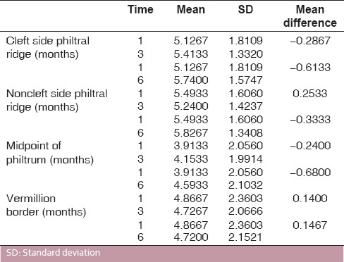
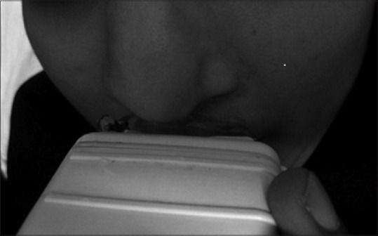
Materials and methods: The study was conducted in SDM Craniofacial Unit, Dharwad and was approved by Local Institutional Review Board. A total of 30 patients, age group ranging from 4 to 25 years, of which 15 postoperative unilateral cleft lip constituted the test group. The remaining 15 with no cleft deformities, no gross facial asymmetry, constituted the control group. The thickness of the mucosa, submucosa, muscle and full thickness of the upper lip were measured with the transversal images using ultrasonography at midpoint of philtrum, right and left side philtral ridges and vermillion border, at 1, 3, 6 months interval.
Results: There was an increase in muscle thickness at the vermillion border (mean = 6.9 mm) and philtral ridge (5.9 mm). Equal muscle thickness were found between the normal and test group at 6 months follow-up in a relaxed position, which was statistically significant (P = 0.0404).
Conclusion: Quantitative assessment of thickness and echo levels of various lip tissues are done with proper echographic calibration. Diagnostic potentials of this method for noninvasive evaluation of cleft lip reconstructions were achieved by this study.


 分享
分享

 求助内容:
求助内容: 应助结果提醒方式:
 扫码关注我们
