Ryan P Goff, Brian T Howard, Stephen G Quallich, Tinen L Iles, Paul A Iaizzo
{"title":"分离的人类和大型哺乳动物心肺块的新颖体外再生。","authors":"Ryan P Goff, Brian T Howard, Stephen G Quallich, Tinen L Iles, Paul A Iaizzo","doi":"10.1186/s12899-016-0023-2","DOIUrl":null,"url":null,"abstract":"<p><strong>Background: </strong>In vitro isolated heart preparations are valuable tools for the study of cardiac anatomy and physiology, as well as for preclinical device testing. Such preparations afford investigators a high level of hemodynamic control, independent of host or systemic interactions. Here we hypothesize that recovered human and swine heart-lung blocs can be reanimated using a clear perfusate and elicit viable cardiodynamic and pulmonic function. Further, this approach will facilitate multimodal imaging, which is particularly valuable for the study of both functional anatomy and device-tissue interactions. Five human and 18 swine heart-lung preparations were procured using techniques analogous to those for cardiac transplant. Specimens were then rewarmed and reperfused using modifications of a closed circuit, isolated, beating and ventilated heart-lung preparation. Positive pressure mechanical ventilation was also employed, and epicardial defibrillation was applied to elicit native cardiac sinus rhythm. Videoscopy, fluoroscopy, ultrasound, and infrared imaging were performed for anatomical and experimental study.</p><p><strong>Results: </strong>Systolic and diastolic left ventricular pressures observed for human and swine specimens were 68/2 ± 11/7 and 74/3 ± 17/5 mmHg, respectively, with associated native heart rates of 80 ± 7 and 96 ± 16 beats per minute. High-resolution imaging within functioning human pulmonary vasculature was obtained among other anatomies of interest. Note that one human specimen elicited poor cardiac performance post defibrillation.</p><p><strong>Conclusions: </strong>We report the first dynamic videoscopic images of the pulmonary vasculature during viable cardiopulmonary function in isolated reanimated heart-lung blocs. This experimental approach provides unique in vitro opportunities for the study of novel medical therapeutics applied to large mammalian, including human, heart-lung specimens.</p>","PeriodicalId":35905,"journal":{"name":"BMC Physiology","volume":"16 1","pages":"4"},"PeriodicalIF":0.0000,"publicationDate":"2016-06-04","publicationTypes":"Journal Article","fieldsOfStudy":null,"isOpenAccess":false,"openAccessPdf":"https://sci-hub-pdf.com/10.1186/s12899-016-0023-2","citationCount":"7","resultStr":"{\"title\":\"The novel in vitro reanimation of isolated human and large mammalian heart-lung blocs.\",\"authors\":\"Ryan P Goff, Brian T Howard, Stephen G Quallich, Tinen L Iles, Paul A Iaizzo\",\"doi\":\"10.1186/s12899-016-0023-2\",\"DOIUrl\":null,\"url\":null,\"abstract\":\"<p><strong>Background: </strong>In vitro isolated heart preparations are valuable tools for the study of cardiac anatomy and physiology, as well as for preclinical device testing. Such preparations afford investigators a high level of hemodynamic control, independent of host or systemic interactions. Here we hypothesize that recovered human and swine heart-lung blocs can be reanimated using a clear perfusate and elicit viable cardiodynamic and pulmonic function. Further, this approach will facilitate multimodal imaging, which is particularly valuable for the study of both functional anatomy and device-tissue interactions. Five human and 18 swine heart-lung preparations were procured using techniques analogous to those for cardiac transplant. Specimens were then rewarmed and reperfused using modifications of a closed circuit, isolated, beating and ventilated heart-lung preparation. Positive pressure mechanical ventilation was also employed, and epicardial defibrillation was applied to elicit native cardiac sinus rhythm. Videoscopy, fluoroscopy, ultrasound, and infrared imaging were performed for anatomical and experimental study.</p><p><strong>Results: </strong>Systolic and diastolic left ventricular pressures observed for human and swine specimens were 68/2 ± 11/7 and 74/3 ± 17/5 mmHg, respectively, with associated native heart rates of 80 ± 7 and 96 ± 16 beats per minute. High-resolution imaging within functioning human pulmonary vasculature was obtained among other anatomies of interest. Note that one human specimen elicited poor cardiac performance post defibrillation.</p><p><strong>Conclusions: </strong>We report the first dynamic videoscopic images of the pulmonary vasculature during viable cardiopulmonary function in isolated reanimated heart-lung blocs. This experimental approach provides unique in vitro opportunities for the study of novel medical therapeutics applied to large mammalian, including human, heart-lung specimens.</p>\",\"PeriodicalId\":35905,\"journal\":{\"name\":\"BMC Physiology\",\"volume\":\"16 1\",\"pages\":\"4\"},\"PeriodicalIF\":0.0000,\"publicationDate\":\"2016-06-04\",\"publicationTypes\":\"Journal Article\",\"fieldsOfStudy\":null,\"isOpenAccess\":false,\"openAccessPdf\":\"https://sci-hub-pdf.com/10.1186/s12899-016-0023-2\",\"citationCount\":\"7\",\"resultStr\":null,\"platform\":\"Semanticscholar\",\"paperid\":null,\"PeriodicalName\":\"BMC Physiology\",\"FirstCategoryId\":\"1085\",\"ListUrlMain\":\"https://doi.org/10.1186/s12899-016-0023-2\",\"RegionNum\":0,\"RegionCategory\":null,\"ArticlePicture\":[],\"TitleCN\":null,\"AbstractTextCN\":null,\"PMCID\":null,\"EPubDate\":\"\",\"PubModel\":\"\",\"JCR\":\"Q1\",\"JCRName\":\"Biochemistry, Genetics and Molecular Biology\",\"Score\":null,\"Total\":0}","platform":"Semanticscholar","paperid":null,"PeriodicalName":"BMC Physiology","FirstCategoryId":"1085","ListUrlMain":"https://doi.org/10.1186/s12899-016-0023-2","RegionNum":0,"RegionCategory":null,"ArticlePicture":[],"TitleCN":null,"AbstractTextCN":null,"PMCID":null,"EPubDate":"","PubModel":"","JCR":"Q1","JCRName":"Biochemistry, Genetics and Molecular Biology","Score":null,"Total":0}
The novel in vitro reanimation of isolated human and large mammalian heart-lung blocs.
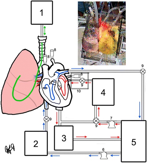
Background: In vitro isolated heart preparations are valuable tools for the study of cardiac anatomy and physiology, as well as for preclinical device testing. Such preparations afford investigators a high level of hemodynamic control, independent of host or systemic interactions. Here we hypothesize that recovered human and swine heart-lung blocs can be reanimated using a clear perfusate and elicit viable cardiodynamic and pulmonic function. Further, this approach will facilitate multimodal imaging, which is particularly valuable for the study of both functional anatomy and device-tissue interactions. Five human and 18 swine heart-lung preparations were procured using techniques analogous to those for cardiac transplant. Specimens were then rewarmed and reperfused using modifications of a closed circuit, isolated, beating and ventilated heart-lung preparation. Positive pressure mechanical ventilation was also employed, and epicardial defibrillation was applied to elicit native cardiac sinus rhythm. Videoscopy, fluoroscopy, ultrasound, and infrared imaging were performed for anatomical and experimental study.
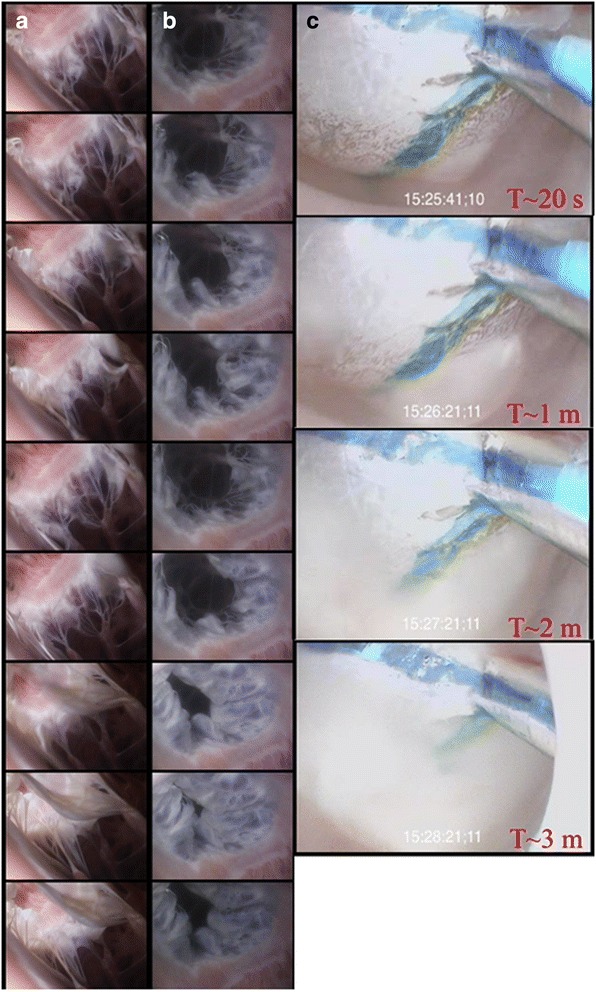
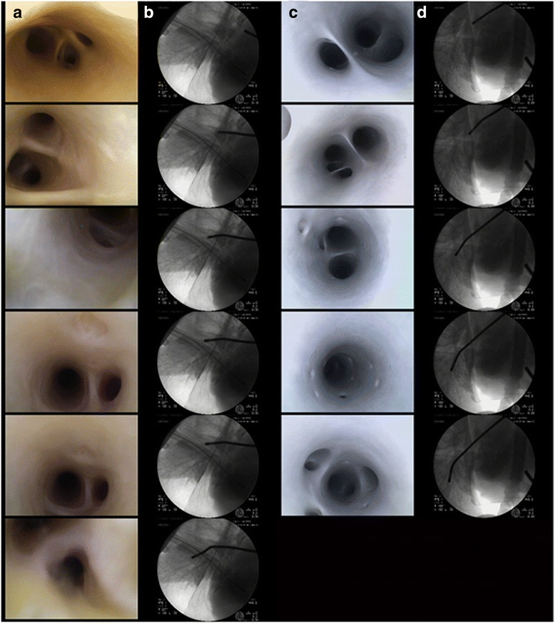
Results: Systolic and diastolic left ventricular pressures observed for human and swine specimens were 68/2 ± 11/7 and 74/3 ± 17/5 mmHg, respectively, with associated native heart rates of 80 ± 7 and 96 ± 16 beats per minute. High-resolution imaging within functioning human pulmonary vasculature was obtained among other anatomies of interest. Note that one human specimen elicited poor cardiac performance post defibrillation.
Conclusions: We report the first dynamic videoscopic images of the pulmonary vasculature during viable cardiopulmonary function in isolated reanimated heart-lung blocs. This experimental approach provides unique in vitro opportunities for the study of novel medical therapeutics applied to large mammalian, including human, heart-lung specimens.
BMC PhysiologyBiochemistry, Genetics and Molecular Biology-Physiology
CiteScore
9.60
自引率
0.00%
发文量
0
期刊介绍:
BMC Physiology is an open access journal publishing original peer-reviewed research articles in cellular, tissue-level, organismal, functional, and developmental aspects of physiological processes. BMC Physiology (ISSN 1472-6793) is indexed/tracked/covered by PubMed, MEDLINE, BIOSIS, CAS, EMBASE, Scopus, Zoological Record and Google Scholar.


 分享
分享
 求助内容:
求助内容: 应助结果提醒方式:
 扫码关注我们
