{"title":"利用数字全景放射摄影的线性和角度测量早期预测下颌第三磨牙萌出/嵌塞:一项放射学研究。","authors":"Rachninder Kaur, Anand C Kumar, Ranjana Garg, Sugandha Sharma, Trisha Rastogi, Vivek Vijay Gupta","doi":"10.4103/0975-962X.184644","DOIUrl":null,"url":null,"abstract":"Background: The impaction rate is higher for the third molars than for any other tooth in modern human population. This study was conducted with the aim to evaluate the validity of linear and angular measurements on the digital panoramic radiograph as a reference for early prediction of mandibular third molar eruption/impaction. Materials and Methods: Digital panoramic radiographs of 200 subjects were selected based on their status of eruption of mandibular third molars; fully erupted (Group A), partially erupted (Group B), fully developed but not erupted (Group C) and partially developed groups (Group D). Each group comprised 50 subjects with 25 males and 25 females. Nine variables (linear measurements, angles, and ratios) were determined and measured bilaterally by two observers and values were compared between the study groups and genders. Results: The data thus obtained were analyzed for comparison among all the study groups. It was found that the difference in the mean values of lower eruption space (LES) measurements, α-angle (angle between long axis of the third molar and gonial-symphyseal plane) and β-angle (angle between long axis of mandibular second and third molars) were significant (P < 0.05). The mean values of mesiodistal width, LES-ramus, LES-Xi point and β-angle were found more in males than in females. No significant difference was observed between the sides. Conclusion: α- and β-angle together with LES measurements give the accurate information on early prediction of lower third molar eruption or impaction.","PeriodicalId":90526,"journal":{"name":"Indian journal of dentistry","volume":"7 2","pages":"66-9"},"PeriodicalIF":0.0000,"publicationDate":"2016-04-01","publicationTypes":"Journal Article","fieldsOfStudy":null,"isOpenAccess":false,"openAccessPdf":"https://ftp.ncbi.nlm.nih.gov/pub/pmc/oa_pdf/e6/16/IJDENT-7-66.PMC4934090.pdf","citationCount":"10","resultStr":"{\"title\":\"Early prediction of mandibular third molar eruption/impaction using linear and angular measurements on digital panoramic radiography: A radiographic study.\",\"authors\":\"Rachninder Kaur, Anand C Kumar, Ranjana Garg, Sugandha Sharma, Trisha Rastogi, Vivek Vijay Gupta\",\"doi\":\"10.4103/0975-962X.184644\",\"DOIUrl\":null,\"url\":null,\"abstract\":\"Background: The impaction rate is higher for the third molars than for any other tooth in modern human population. This study was conducted with the aim to evaluate the validity of linear and angular measurements on the digital panoramic radiograph as a reference for early prediction of mandibular third molar eruption/impaction. Materials and Methods: Digital panoramic radiographs of 200 subjects were selected based on their status of eruption of mandibular third molars; fully erupted (Group A), partially erupted (Group B), fully developed but not erupted (Group C) and partially developed groups (Group D). Each group comprised 50 subjects with 25 males and 25 females. Nine variables (linear measurements, angles, and ratios) were determined and measured bilaterally by two observers and values were compared between the study groups and genders. Results: The data thus obtained were analyzed for comparison among all the study groups. It was found that the difference in the mean values of lower eruption space (LES) measurements, α-angle (angle between long axis of the third molar and gonial-symphyseal plane) and β-angle (angle between long axis of mandibular second and third molars) were significant (P < 0.05). The mean values of mesiodistal width, LES-ramus, LES-Xi point and β-angle were found more in males than in females. No significant difference was observed between the sides. Conclusion: α- and β-angle together with LES measurements give the accurate information on early prediction of lower third molar eruption or impaction.\",\"PeriodicalId\":90526,\"journal\":{\"name\":\"Indian journal of dentistry\",\"volume\":\"7 2\",\"pages\":\"66-9\"},\"PeriodicalIF\":0.0000,\"publicationDate\":\"2016-04-01\",\"publicationTypes\":\"Journal Article\",\"fieldsOfStudy\":null,\"isOpenAccess\":false,\"openAccessPdf\":\"https://ftp.ncbi.nlm.nih.gov/pub/pmc/oa_pdf/e6/16/IJDENT-7-66.PMC4934090.pdf\",\"citationCount\":\"10\",\"resultStr\":null,\"platform\":\"Semanticscholar\",\"paperid\":null,\"PeriodicalName\":\"Indian journal of dentistry\",\"FirstCategoryId\":\"1085\",\"ListUrlMain\":\"https://doi.org/10.4103/0975-962X.184644\",\"RegionNum\":0,\"RegionCategory\":null,\"ArticlePicture\":[],\"TitleCN\":null,\"AbstractTextCN\":null,\"PMCID\":null,\"EPubDate\":\"\",\"PubModel\":\"\",\"JCR\":\"\",\"JCRName\":\"\",\"Score\":null,\"Total\":0}","platform":"Semanticscholar","paperid":null,"PeriodicalName":"Indian journal of dentistry","FirstCategoryId":"1085","ListUrlMain":"https://doi.org/10.4103/0975-962X.184644","RegionNum":0,"RegionCategory":null,"ArticlePicture":[],"TitleCN":null,"AbstractTextCN":null,"PMCID":null,"EPubDate":"","PubModel":"","JCR":"","JCRName":"","Score":null,"Total":0}
Early prediction of mandibular third molar eruption/impaction using linear and angular measurements on digital panoramic radiography: A radiographic study.
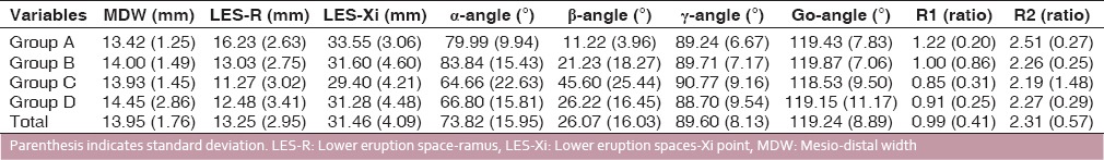
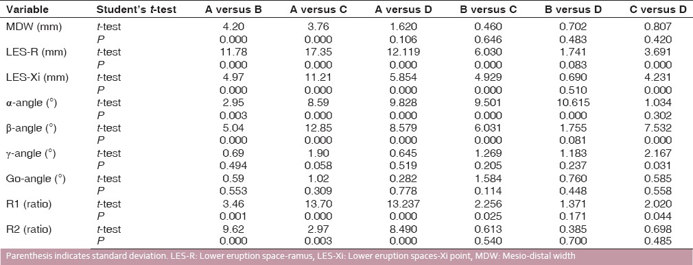
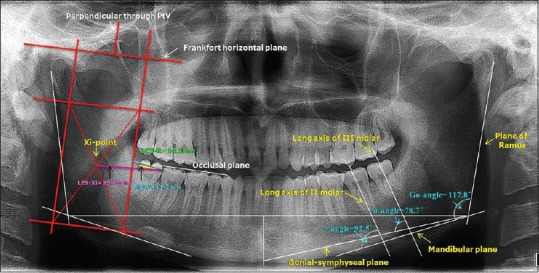
Background: The impaction rate is higher for the third molars than for any other tooth in modern human population. This study was conducted with the aim to evaluate the validity of linear and angular measurements on the digital panoramic radiograph as a reference for early prediction of mandibular third molar eruption/impaction. Materials and Methods: Digital panoramic radiographs of 200 subjects were selected based on their status of eruption of mandibular third molars; fully erupted (Group A), partially erupted (Group B), fully developed but not erupted (Group C) and partially developed groups (Group D). Each group comprised 50 subjects with 25 males and 25 females. Nine variables (linear measurements, angles, and ratios) were determined and measured bilaterally by two observers and values were compared between the study groups and genders. Results: The data thus obtained were analyzed for comparison among all the study groups. It was found that the difference in the mean values of lower eruption space (LES) measurements, α-angle (angle between long axis of the third molar and gonial-symphyseal plane) and β-angle (angle between long axis of mandibular second and third molars) were significant (P < 0.05). The mean values of mesiodistal width, LES-ramus, LES-Xi point and β-angle were found more in males than in females. No significant difference was observed between the sides. Conclusion: α- and β-angle together with LES measurements give the accurate information on early prediction of lower third molar eruption or impaction.


 分享
分享

 求助内容:
求助内容: 应助结果提醒方式:
 扫码关注我们
