{"title":"伴有腔腔和壁腔成分的钙化牙源性囊肿(1c型)。","authors":"Bhushan Sharma, George Koshy, Shekhar Kapoor","doi":"10.4103/0975-962X.184648","DOIUrl":null,"url":null,"abstract":"<p><p>Calcifying odontogenic cyst (COC) was first described and classified by Gorlin et al. It is defined as a cystic lesion in which the epithelial lining shows a well defined basal layer of columnar cells, an overlying layer that often resemble stellate reticulum and masses of ghost cells that may be in the epithelial cystic lining or in the fibrous capsule. The lesion generally occurs in the region anterior to maxillary and mandibular molars and either intraosseous or extraosseus. This entity might present as a cystic or solid lesion. Praetorius et al. classified COC into 2 main entities namely a cyst (Type 1) and a neoplasm (Type 2). The present case report exhibit a cystic lesion with both luminal and mural component. </p>","PeriodicalId":90526,"journal":{"name":"Indian journal of dentistry","volume":"7 2","pages":"95-8"},"PeriodicalIF":0.0000,"publicationDate":"2016-04-01","publicationTypes":"Journal Article","fieldsOfStudy":null,"isOpenAccess":false,"openAccessPdf":"https://ftp.ncbi.nlm.nih.gov/pub/pmc/oa_pdf/6d/89/IJDENT-7-95.PMC4934095.pdf","citationCount":"3","resultStr":"{\"title\":\"Calcifying odontogenic cyst with luminal and mural component (Type 1c).\",\"authors\":\"Bhushan Sharma, George Koshy, Shekhar Kapoor\",\"doi\":\"10.4103/0975-962X.184648\",\"DOIUrl\":null,\"url\":null,\"abstract\":\"<p><p>Calcifying odontogenic cyst (COC) was first described and classified by Gorlin et al. It is defined as a cystic lesion in which the epithelial lining shows a well defined basal layer of columnar cells, an overlying layer that often resemble stellate reticulum and masses of ghost cells that may be in the epithelial cystic lining or in the fibrous capsule. The lesion generally occurs in the region anterior to maxillary and mandibular molars and either intraosseous or extraosseus. This entity might present as a cystic or solid lesion. Praetorius et al. classified COC into 2 main entities namely a cyst (Type 1) and a neoplasm (Type 2). The present case report exhibit a cystic lesion with both luminal and mural component. </p>\",\"PeriodicalId\":90526,\"journal\":{\"name\":\"Indian journal of dentistry\",\"volume\":\"7 2\",\"pages\":\"95-8\"},\"PeriodicalIF\":0.0000,\"publicationDate\":\"2016-04-01\",\"publicationTypes\":\"Journal Article\",\"fieldsOfStudy\":null,\"isOpenAccess\":false,\"openAccessPdf\":\"https://ftp.ncbi.nlm.nih.gov/pub/pmc/oa_pdf/6d/89/IJDENT-7-95.PMC4934095.pdf\",\"citationCount\":\"3\",\"resultStr\":null,\"platform\":\"Semanticscholar\",\"paperid\":null,\"PeriodicalName\":\"Indian journal of dentistry\",\"FirstCategoryId\":\"1085\",\"ListUrlMain\":\"https://doi.org/10.4103/0975-962X.184648\",\"RegionNum\":0,\"RegionCategory\":null,\"ArticlePicture\":[],\"TitleCN\":null,\"AbstractTextCN\":null,\"PMCID\":null,\"EPubDate\":\"\",\"PubModel\":\"\",\"JCR\":\"\",\"JCRName\":\"\",\"Score\":null,\"Total\":0}","platform":"Semanticscholar","paperid":null,"PeriodicalName":"Indian journal of dentistry","FirstCategoryId":"1085","ListUrlMain":"https://doi.org/10.4103/0975-962X.184648","RegionNum":0,"RegionCategory":null,"ArticlePicture":[],"TitleCN":null,"AbstractTextCN":null,"PMCID":null,"EPubDate":"","PubModel":"","JCR":"","JCRName":"","Score":null,"Total":0}
Calcifying odontogenic cyst with luminal and mural component (Type 1c).
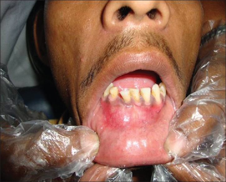
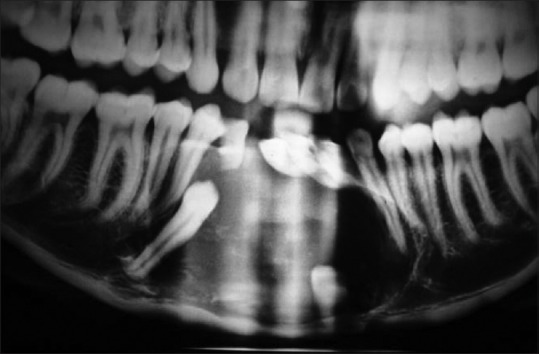
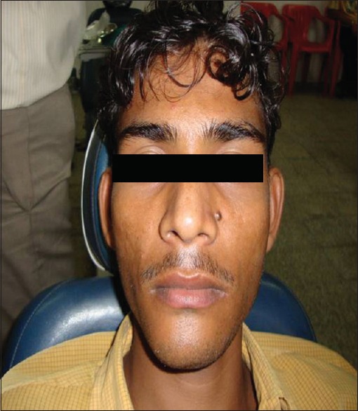
Calcifying odontogenic cyst (COC) was first described and classified by Gorlin et al. It is defined as a cystic lesion in which the epithelial lining shows a well defined basal layer of columnar cells, an overlying layer that often resemble stellate reticulum and masses of ghost cells that may be in the epithelial cystic lining or in the fibrous capsule. The lesion generally occurs in the region anterior to maxillary and mandibular molars and either intraosseous or extraosseus. This entity might present as a cystic or solid lesion. Praetorius et al. classified COC into 2 main entities namely a cyst (Type 1) and a neoplasm (Type 2). The present case report exhibit a cystic lesion with both luminal and mural component.


 分享
分享

 求助内容:
求助内容: 应助结果提醒方式:
 扫码关注我们
