{"title":"IN/OUT试验:研究纤毛发生的新工具。","authors":"Ira Kukic, Felix Rivera-Molina, Derek Toomre","doi":"10.1186/s13630-016-0044-2","DOIUrl":null,"url":null,"abstract":"<p><strong>Background: </strong>Nearly all cells have a primary cilia on their surface, which functions as a cellular antennae. Primary cilia assembly begins intracellularly and eventually emerges extracellularly. However, current ciliogenesis assays, which detect cilia length and number, do not monitor ciliary stages.</p><p><strong>Methods: </strong>We developed a new assay that detects antibody access to a fluorescently tagged ciliary transmembrane protein, which revealed three ciliary states: classified as 'inside,' 'outside,' or 'partial' cilia.</p><p><strong>Results: </strong>Strikingly, most cilia in RPE cells only partially emerged and many others were long and intracellular, which would be indistinguishable by conventional assays. Importantly, these states switch with starvation-induced ciliogenesis and the cilia can emerge both on the dorsal and ventral surface of the cell. Our assay further allows new molecular and functional studies of the 'ciliary pocket,' a deep plasma membrane invagination whose function is unclear. Molecularly, we show colocalization of EHD1, Septin 9 and glutamylated tubulin with the ciliary pocket.</p><p><strong>Conclusions: </strong>Together, the IN/OUT assay is not only a new tool for easy and quantifiable visualization of different ciliary stages, but also allows molecular characterization of intermediate ciliary states.</p>","PeriodicalId":38134,"journal":{"name":"Cilia","volume":"5 ","pages":"23"},"PeriodicalIF":0.0000,"publicationDate":"2016-08-04","publicationTypes":"Journal Article","fieldsOfStudy":null,"isOpenAccess":false,"openAccessPdf":"https://sci-hub-pdf.com/10.1186/s13630-016-0044-2","citationCount":"34","resultStr":"{\"title\":\"The IN/OUT assay: a new tool to study ciliogenesis.\",\"authors\":\"Ira Kukic, Felix Rivera-Molina, Derek Toomre\",\"doi\":\"10.1186/s13630-016-0044-2\",\"DOIUrl\":null,\"url\":null,\"abstract\":\"<p><strong>Background: </strong>Nearly all cells have a primary cilia on their surface, which functions as a cellular antennae. Primary cilia assembly begins intracellularly and eventually emerges extracellularly. However, current ciliogenesis assays, which detect cilia length and number, do not monitor ciliary stages.</p><p><strong>Methods: </strong>We developed a new assay that detects antibody access to a fluorescently tagged ciliary transmembrane protein, which revealed three ciliary states: classified as 'inside,' 'outside,' or 'partial' cilia.</p><p><strong>Results: </strong>Strikingly, most cilia in RPE cells only partially emerged and many others were long and intracellular, which would be indistinguishable by conventional assays. Importantly, these states switch with starvation-induced ciliogenesis and the cilia can emerge both on the dorsal and ventral surface of the cell. Our assay further allows new molecular and functional studies of the 'ciliary pocket,' a deep plasma membrane invagination whose function is unclear. Molecularly, we show colocalization of EHD1, Septin 9 and glutamylated tubulin with the ciliary pocket.</p><p><strong>Conclusions: </strong>Together, the IN/OUT assay is not only a new tool for easy and quantifiable visualization of different ciliary stages, but also allows molecular characterization of intermediate ciliary states.</p>\",\"PeriodicalId\":38134,\"journal\":{\"name\":\"Cilia\",\"volume\":\"5 \",\"pages\":\"23\"},\"PeriodicalIF\":0.0000,\"publicationDate\":\"2016-08-04\",\"publicationTypes\":\"Journal Article\",\"fieldsOfStudy\":null,\"isOpenAccess\":false,\"openAccessPdf\":\"https://sci-hub-pdf.com/10.1186/s13630-016-0044-2\",\"citationCount\":\"34\",\"resultStr\":null,\"platform\":\"Semanticscholar\",\"paperid\":null,\"PeriodicalName\":\"Cilia\",\"FirstCategoryId\":\"1085\",\"ListUrlMain\":\"https://doi.org/10.1186/s13630-016-0044-2\",\"RegionNum\":0,\"RegionCategory\":null,\"ArticlePicture\":[],\"TitleCN\":null,\"AbstractTextCN\":null,\"PMCID\":null,\"EPubDate\":\"2016/1/1 0:00:00\",\"PubModel\":\"eCollection\",\"JCR\":\"Q2\",\"JCRName\":\"Biochemistry, Genetics and Molecular Biology\",\"Score\":null,\"Total\":0}","platform":"Semanticscholar","paperid":null,"PeriodicalName":"Cilia","FirstCategoryId":"1085","ListUrlMain":"https://doi.org/10.1186/s13630-016-0044-2","RegionNum":0,"RegionCategory":null,"ArticlePicture":[],"TitleCN":null,"AbstractTextCN":null,"PMCID":null,"EPubDate":"2016/1/1 0:00:00","PubModel":"eCollection","JCR":"Q2","JCRName":"Biochemistry, Genetics and Molecular Biology","Score":null,"Total":0}
The IN/OUT assay: a new tool to study ciliogenesis.
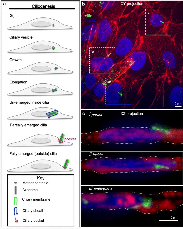
Background: Nearly all cells have a primary cilia on their surface, which functions as a cellular antennae. Primary cilia assembly begins intracellularly and eventually emerges extracellularly. However, current ciliogenesis assays, which detect cilia length and number, do not monitor ciliary stages.
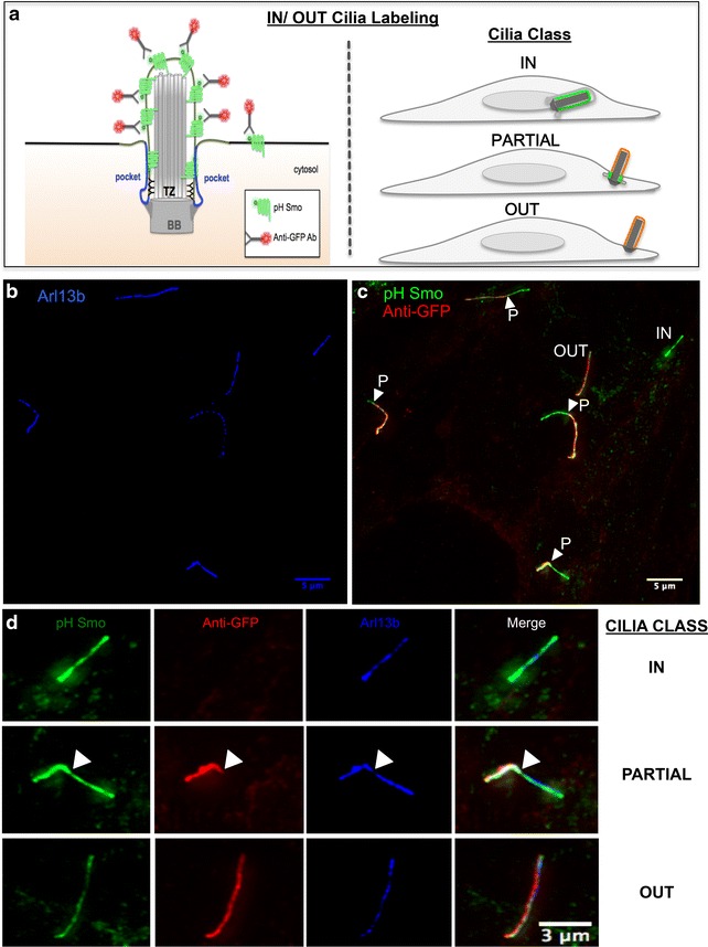
Methods: We developed a new assay that detects antibody access to a fluorescently tagged ciliary transmembrane protein, which revealed three ciliary states: classified as 'inside,' 'outside,' or 'partial' cilia.
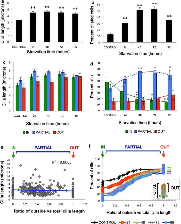
Results: Strikingly, most cilia in RPE cells only partially emerged and many others were long and intracellular, which would be indistinguishable by conventional assays. Importantly, these states switch with starvation-induced ciliogenesis and the cilia can emerge both on the dorsal and ventral surface of the cell. Our assay further allows new molecular and functional studies of the 'ciliary pocket,' a deep plasma membrane invagination whose function is unclear. Molecularly, we show colocalization of EHD1, Septin 9 and glutamylated tubulin with the ciliary pocket.
Conclusions: Together, the IN/OUT assay is not only a new tool for easy and quantifiable visualization of different ciliary stages, but also allows molecular characterization of intermediate ciliary states.


 分享
分享
 求助内容:
求助内容: 应助结果提醒方式:
 扫码关注我们
