Emre Demirci, Rafay Ahmed, Meltem Ocak, Joseph Latoche, April Radelet, Nicole DeBlasio, N Scott Mason, Carolyn J Anderson, James M Mountz
{"title":"18F-ML-10的临床前评估以确定实体肿瘤对化疗的凋亡反应时间。","authors":"Emre Demirci, Rafay Ahmed, Meltem Ocak, Joseph Latoche, April Radelet, Nicole DeBlasio, N Scott Mason, Carolyn J Anderson, James M Mountz","doi":"10.1177/1536012116685941","DOIUrl":null,"url":null,"abstract":"<p><strong>Purpose: </strong>We investigated 2-(5-fluoro-pentyl)-2-methyl-malonic acid (<sup>18</sup>F-ML-10) positron emission tomography (PET) imaging of apoptosis posttherapy to determine optimal timing for predicting chemotherapy response in a mouse head/neck xenograft cancer model.</p><p><strong>Procedures: </strong>BALB/c nude mice (4-8 weeks old) were implanted with UM-SCC-22B tumors. The treatment group received 2 doses of doxorubicin (10 mg/kg, days 0, 2). Small animal <sup>18</sup>F-ML-10 PET/computed tomography was performed before and on days 1, 3, and 7 postchemotherapy. Using regions of interest around tumors, <sup>18</sup>F-ML-10 uptake change was measured as %ID/g and uptake relative to liver. Terminal Uridine Nick-End Labeling (TUNEL) immunohistochemistry assay was performed using tumor samples of baseline and on days 1, 3, and 7 posttreatment.</p><p><strong>Results: </strong>Treated mice demonstrated increased <sup>18</sup>F-ML-10 uptake compared to baseline and controls, and 10 of 13 mice showed tumor volume decreases. All control mice showed tumor volume increases. Tumor-to-liver (T/L) ratios from the control group mice did not show significant change from baseline ( P > .05); however, T/L ratios of the treatment group showed significant <sup>18</sup>F-ML-10 uptake differences from baseline compared to days 3 and 7 posttreatment ( P < .05), but no significant difference at 1 day posttreatment.</p><p><strong>Conclusion: </strong>2-(5-Fluoro-pentyl)-2-methyl-malonic acid PET imaging has the potential for early assessment of treatment-induced apoptosis. Timing and image analysis strategies may require optimization, depending on the type of tumor and cancer treatment.</p>","PeriodicalId":18855,"journal":{"name":"Molecular Imaging","volume":"16 ","pages":"1536012116685941"},"PeriodicalIF":2.4000,"publicationDate":"2017-01-01","publicationTypes":"Journal Article","fieldsOfStudy":null,"isOpenAccess":false,"openAccessPdf":"https://sci-hub-pdf.com/10.1177/1536012116685941","citationCount":"14","resultStr":"{\"title\":\"Preclinical Evaluation of <sup>18</sup>F-ML-10 to Determine Timing of Apoptotic Response to Chemotherapy in Solid Tumors.\",\"authors\":\"Emre Demirci, Rafay Ahmed, Meltem Ocak, Joseph Latoche, April Radelet, Nicole DeBlasio, N Scott Mason, Carolyn J Anderson, James M Mountz\",\"doi\":\"10.1177/1536012116685941\",\"DOIUrl\":null,\"url\":null,\"abstract\":\"<p><strong>Purpose: </strong>We investigated 2-(5-fluoro-pentyl)-2-methyl-malonic acid (<sup>18</sup>F-ML-10) positron emission tomography (PET) imaging of apoptosis posttherapy to determine optimal timing for predicting chemotherapy response in a mouse head/neck xenograft cancer model.</p><p><strong>Procedures: </strong>BALB/c nude mice (4-8 weeks old) were implanted with UM-SCC-22B tumors. The treatment group received 2 doses of doxorubicin (10 mg/kg, days 0, 2). Small animal <sup>18</sup>F-ML-10 PET/computed tomography was performed before and on days 1, 3, and 7 postchemotherapy. Using regions of interest around tumors, <sup>18</sup>F-ML-10 uptake change was measured as %ID/g and uptake relative to liver. Terminal Uridine Nick-End Labeling (TUNEL) immunohistochemistry assay was performed using tumor samples of baseline and on days 1, 3, and 7 posttreatment.</p><p><strong>Results: </strong>Treated mice demonstrated increased <sup>18</sup>F-ML-10 uptake compared to baseline and controls, and 10 of 13 mice showed tumor volume decreases. All control mice showed tumor volume increases. Tumor-to-liver (T/L) ratios from the control group mice did not show significant change from baseline ( P > .05); however, T/L ratios of the treatment group showed significant <sup>18</sup>F-ML-10 uptake differences from baseline compared to days 3 and 7 posttreatment ( P < .05), but no significant difference at 1 day posttreatment.</p><p><strong>Conclusion: </strong>2-(5-Fluoro-pentyl)-2-methyl-malonic acid PET imaging has the potential for early assessment of treatment-induced apoptosis. Timing and image analysis strategies may require optimization, depending on the type of tumor and cancer treatment.</p>\",\"PeriodicalId\":18855,\"journal\":{\"name\":\"Molecular Imaging\",\"volume\":\"16 \",\"pages\":\"1536012116685941\"},\"PeriodicalIF\":2.4000,\"publicationDate\":\"2017-01-01\",\"publicationTypes\":\"Journal Article\",\"fieldsOfStudy\":null,\"isOpenAccess\":false,\"openAccessPdf\":\"https://sci-hub-pdf.com/10.1177/1536012116685941\",\"citationCount\":\"14\",\"resultStr\":null,\"platform\":\"Semanticscholar\",\"paperid\":null,\"PeriodicalName\":\"Molecular Imaging\",\"FirstCategoryId\":\"3\",\"ListUrlMain\":\"https://doi.org/10.1177/1536012116685941\",\"RegionNum\":4,\"RegionCategory\":\"医学\",\"ArticlePicture\":[],\"TitleCN\":null,\"AbstractTextCN\":null,\"PMCID\":null,\"EPubDate\":\"\",\"PubModel\":\"\",\"JCR\":\"Q3\",\"JCRName\":\"BIOCHEMICAL RESEARCH METHODS\",\"Score\":null,\"Total\":0}","platform":"Semanticscholar","paperid":null,"PeriodicalName":"Molecular Imaging","FirstCategoryId":"3","ListUrlMain":"https://doi.org/10.1177/1536012116685941","RegionNum":4,"RegionCategory":"医学","ArticlePicture":[],"TitleCN":null,"AbstractTextCN":null,"PMCID":null,"EPubDate":"","PubModel":"","JCR":"Q3","JCRName":"BIOCHEMICAL RESEARCH METHODS","Score":null,"Total":0}
Preclinical Evaluation of 18F-ML-10 to Determine Timing of Apoptotic Response to Chemotherapy in Solid Tumors.
Purpose: We investigated 2-(5-fluoro-pentyl)-2-methyl-malonic acid (18F-ML-10) positron emission tomography (PET) imaging of apoptosis posttherapy to determine optimal timing for predicting chemotherapy response in a mouse head/neck xenograft cancer model.
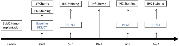
Procedures: BALB/c nude mice (4-8 weeks old) were implanted with UM-SCC-22B tumors. The treatment group received 2 doses of doxorubicin (10 mg/kg, days 0, 2). Small animal 18F-ML-10 PET/computed tomography was performed before and on days 1, 3, and 7 postchemotherapy. Using regions of interest around tumors, 18F-ML-10 uptake change was measured as %ID/g and uptake relative to liver. Terminal Uridine Nick-End Labeling (TUNEL) immunohistochemistry assay was performed using tumor samples of baseline and on days 1, 3, and 7 posttreatment.
Results: Treated mice demonstrated increased 18F-ML-10 uptake compared to baseline and controls, and 10 of 13 mice showed tumor volume decreases. All control mice showed tumor volume increases. Tumor-to-liver (T/L) ratios from the control group mice did not show significant change from baseline ( P > .05); however, T/L ratios of the treatment group showed significant 18F-ML-10 uptake differences from baseline compared to days 3 and 7 posttreatment ( P < .05), but no significant difference at 1 day posttreatment.
Conclusion: 2-(5-Fluoro-pentyl)-2-methyl-malonic acid PET imaging has the potential for early assessment of treatment-induced apoptosis. Timing and image analysis strategies may require optimization, depending on the type of tumor and cancer treatment.
Molecular ImagingBiochemistry, Genetics and Molecular Biology-Biotechnology
自引率
3.60%
发文量
21
期刊介绍:
Molecular Imaging is a peer-reviewed, open access journal highlighting the breadth of molecular imaging research from basic science to preclinical studies to human applications. This serves both the scientific and clinical communities by disseminating novel results and concepts relevant to the biological study of normal and disease processes in both basic and translational studies ranging from mice to humans.


 分享
分享
 求助内容:
求助内容: 应助结果提醒方式:
 扫码关注我们
