Alexander Krotow, Emine B Yalcin, Jared Kay, Suzanne M de la Monte
{"title":"脂质提取物与成像质谱法评价二手烟暴露实验模型脑白质生化病理的对比分析","authors":"Alexander Krotow, Emine B Yalcin, Jared Kay, Suzanne M de la Monte","doi":"10.4172/2469-9861.1000113","DOIUrl":null,"url":null,"abstract":"<p><strong>Background: </strong>White matter injury and degeneration are common features of developmental and aging-associated diseases, yet their pathobiological bases are poorly understood. However, recent advances in Matrix-Assisted Laser Desorption Ionization (MALDI) instruments and chemistry have provided critical tools for myelin-lipid analytical research.</p><p><strong>Design: </strong>This study characterizes Cigarette Smoke (CS) exposure effects on frontal lobe lipid ion profiles in adult male A/J mice that had been exposed to air for 8 weeks (A8), CS for 4 (CS4) or 8 weeks (CS8), or CS8 followed by 2 weeks recovery (CS8+R). MALDI data acquired by analysis of lipid extracts plated onto a ground steel target (high through-put) were compared with Imaging Mass Spectrometry (IMS).</p><p><strong>Results: </strong>MALDI-time-of-flight (TOF) detected 120 lipid ions with m/z's of 600 to 1300 (phospholipids and sulfatides) in samples plated onto the steel target or analyzed by IMS, but just 25 ions (18%) were detected by both methods. IMS more effectively detected ions in the highest m/z range, whereas the extracts had abundant middle-range m/z ions. The experimental groups were better discriminated by PCA and R-generated heat map hierarchical clustering of IMS data than lipid extract data. On the other hand, both methods clearly delineated the CS4, CS8 and CS8+R experimental groups from control.</p><p><strong>Conclusions: </strong>MALDI analysis of brain lipid extracts plated onto a ground steel target for high through-put studies, or imaged directly in tissue can be used to assess biochemical pathology of white matter neurodegeneration and responses to treatment.</p>","PeriodicalId":92098,"journal":{"name":"Mass spectrometry & purification techniques","volume":"2 1","pages":""},"PeriodicalIF":0.0000,"publicationDate":"2016-01-01","publicationTypes":"Journal Article","fieldsOfStudy":null,"isOpenAccess":false,"openAccessPdf":"https://sci-hub-pdf.com/10.4172/2469-9861.1000113","citationCount":"2","resultStr":"{\"title\":\"Comparative Analysis of Lipid Extracts and Imaging Mass Spectrometry for Evaluating Cerebral White Matter Biochemical Pathology in an Experimental Second-Hand Cigarette Smoke Exposure Model.\",\"authors\":\"Alexander Krotow, Emine B Yalcin, Jared Kay, Suzanne M de la Monte\",\"doi\":\"10.4172/2469-9861.1000113\",\"DOIUrl\":null,\"url\":null,\"abstract\":\"<p><strong>Background: </strong>White matter injury and degeneration are common features of developmental and aging-associated diseases, yet their pathobiological bases are poorly understood. However, recent advances in Matrix-Assisted Laser Desorption Ionization (MALDI) instruments and chemistry have provided critical tools for myelin-lipid analytical research.</p><p><strong>Design: </strong>This study characterizes Cigarette Smoke (CS) exposure effects on frontal lobe lipid ion profiles in adult male A/J mice that had been exposed to air for 8 weeks (A8), CS for 4 (CS4) or 8 weeks (CS8), or CS8 followed by 2 weeks recovery (CS8+R). MALDI data acquired by analysis of lipid extracts plated onto a ground steel target (high through-put) were compared with Imaging Mass Spectrometry (IMS).</p><p><strong>Results: </strong>MALDI-time-of-flight (TOF) detected 120 lipid ions with m/z's of 600 to 1300 (phospholipids and sulfatides) in samples plated onto the steel target or analyzed by IMS, but just 25 ions (18%) were detected by both methods. IMS more effectively detected ions in the highest m/z range, whereas the extracts had abundant middle-range m/z ions. The experimental groups were better discriminated by PCA and R-generated heat map hierarchical clustering of IMS data than lipid extract data. On the other hand, both methods clearly delineated the CS4, CS8 and CS8+R experimental groups from control.</p><p><strong>Conclusions: </strong>MALDI analysis of brain lipid extracts plated onto a ground steel target for high through-put studies, or imaged directly in tissue can be used to assess biochemical pathology of white matter neurodegeneration and responses to treatment.</p>\",\"PeriodicalId\":92098,\"journal\":{\"name\":\"Mass spectrometry & purification techniques\",\"volume\":\"2 1\",\"pages\":\"\"},\"PeriodicalIF\":0.0000,\"publicationDate\":\"2016-01-01\",\"publicationTypes\":\"Journal Article\",\"fieldsOfStudy\":null,\"isOpenAccess\":false,\"openAccessPdf\":\"https://sci-hub-pdf.com/10.4172/2469-9861.1000113\",\"citationCount\":\"2\",\"resultStr\":null,\"platform\":\"Semanticscholar\",\"paperid\":null,\"PeriodicalName\":\"Mass spectrometry & purification techniques\",\"FirstCategoryId\":\"1085\",\"ListUrlMain\":\"https://doi.org/10.4172/2469-9861.1000113\",\"RegionNum\":0,\"RegionCategory\":null,\"ArticlePicture\":[],\"TitleCN\":null,\"AbstractTextCN\":null,\"PMCID\":null,\"EPubDate\":\"2016/4/20 0:00:00\",\"PubModel\":\"Epub\",\"JCR\":\"\",\"JCRName\":\"\",\"Score\":null,\"Total\":0}","platform":"Semanticscholar","paperid":null,"PeriodicalName":"Mass spectrometry & purification techniques","FirstCategoryId":"1085","ListUrlMain":"https://doi.org/10.4172/2469-9861.1000113","RegionNum":0,"RegionCategory":null,"ArticlePicture":[],"TitleCN":null,"AbstractTextCN":null,"PMCID":null,"EPubDate":"2016/4/20 0:00:00","PubModel":"Epub","JCR":"","JCRName":"","Score":null,"Total":0}
Comparative Analysis of Lipid Extracts and Imaging Mass Spectrometry for Evaluating Cerebral White Matter Biochemical Pathology in an Experimental Second-Hand Cigarette Smoke Exposure Model.
Background: White matter injury and degeneration are common features of developmental and aging-associated diseases, yet their pathobiological bases are poorly understood. However, recent advances in Matrix-Assisted Laser Desorption Ionization (MALDI) instruments and chemistry have provided critical tools for myelin-lipid analytical research.
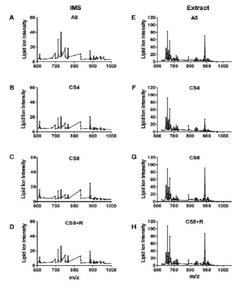
Design: This study characterizes Cigarette Smoke (CS) exposure effects on frontal lobe lipid ion profiles in adult male A/J mice that had been exposed to air for 8 weeks (A8), CS for 4 (CS4) or 8 weeks (CS8), or CS8 followed by 2 weeks recovery (CS8+R). MALDI data acquired by analysis of lipid extracts plated onto a ground steel target (high through-put) were compared with Imaging Mass Spectrometry (IMS).
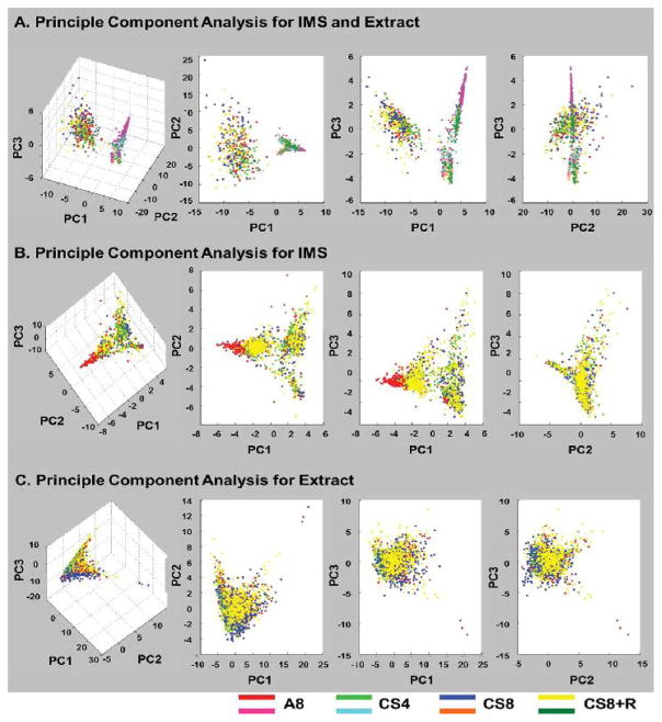
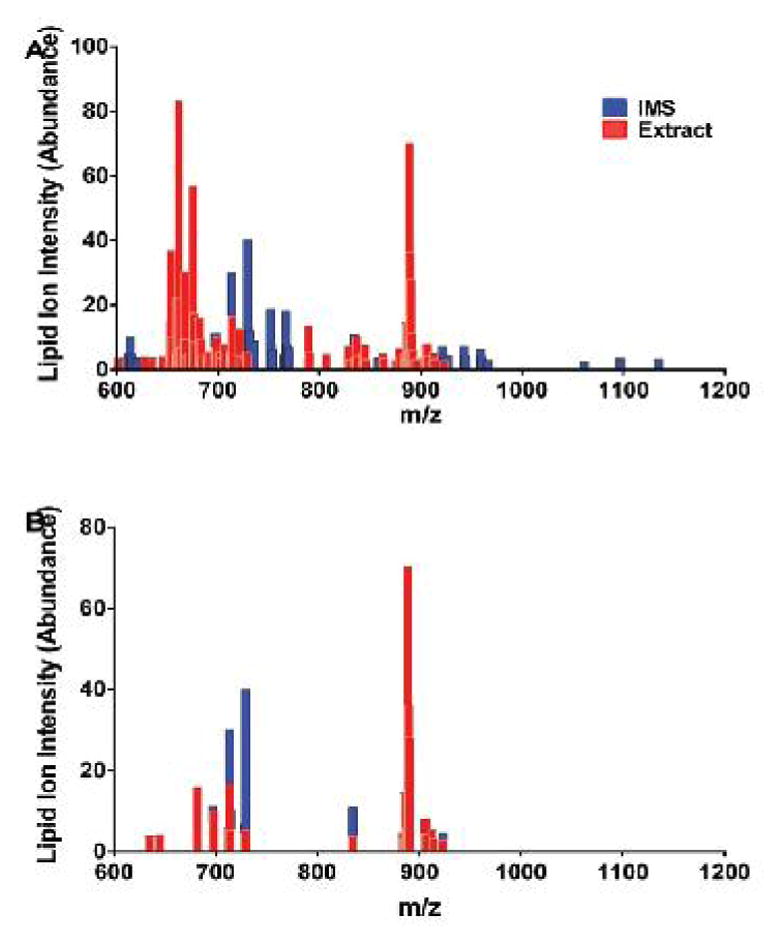
Results: MALDI-time-of-flight (TOF) detected 120 lipid ions with m/z's of 600 to 1300 (phospholipids and sulfatides) in samples plated onto the steel target or analyzed by IMS, but just 25 ions (18%) were detected by both methods. IMS more effectively detected ions in the highest m/z range, whereas the extracts had abundant middle-range m/z ions. The experimental groups were better discriminated by PCA and R-generated heat map hierarchical clustering of IMS data than lipid extract data. On the other hand, both methods clearly delineated the CS4, CS8 and CS8+R experimental groups from control.
Conclusions: MALDI analysis of brain lipid extracts plated onto a ground steel target for high through-put studies, or imaged directly in tissue can be used to assess biochemical pathology of white matter neurodegeneration and responses to treatment.


 分享
分享

 求助内容:
求助内容: 应助结果提醒方式:
 扫码关注我们
