Charlotte Es Hoogstins, Benjamin Weixler, Leonora Sf Boogerd, Diederik J Hoppener, Hendrica Ajm Prevoo, Cornelis Fm Sier, Jacobus Wa Burger, Cornelis Verhoef, Shadvhi Bhairosingh, Arantza Farina Sarasqueta, Jacobus Burggraaf, Alexander L Vahrmeijer
{"title":"寻找结直肠癌腹膜转移术中荧光成像的最佳靶点。","authors":"Charlotte Es Hoogstins, Benjamin Weixler, Leonora Sf Boogerd, Diederik J Hoppener, Hendrica Ajm Prevoo, Cornelis Fm Sier, Jacobus Wa Burger, Cornelis Verhoef, Shadvhi Bhairosingh, Arantza Farina Sarasqueta, Jacobus Burggraaf, Alexander L Vahrmeijer","doi":"10.1177/1179299X17728254","DOIUrl":null,"url":null,"abstract":"<p><p>Peritoneal metastasis (PM) occurs in about 10% of patients with colorectal cancer (CRC). Fluorescence imaging can enhance contrast between cancerous and benign tissue, enabling the surgeon to clearly visualize PM during cytoreductive surgery. This study assessed the suitability of different biomarkers as potential targets for tumor-targeted imaging of PM of CRC. Tissue samples from primary tumor and PM from patients with CRC were obtained from the pathology archives and immunohistochemical stainings were performed. Overexpression of the epithelial cell adhesion molecule (EpCAM) and carcinoembryonic antigen (CEA) was seen in 100% of PM samples and the expression was strong in >70% of samples. Tyrosine-kinase Met (C-Met) and folate receptor α overexpression was seen in 20% of PM samples. For successful application of tumor-targeted intraoperative fluorescence imaging of PM, biomarkers need to be identified. We demonstrated that both EpCAM and CEA are suitable targets for fluorescence imaging of PM in patients with CRC.</p>","PeriodicalId":72377,"journal":{"name":"Biomarkers in cancer","volume":" ","pages":"1179299X17728254"},"PeriodicalIF":0.0000,"publicationDate":"2017-08-28","publicationTypes":"Journal Article","fieldsOfStudy":null,"isOpenAccess":false,"openAccessPdf":"https://sci-hub-pdf.com/10.1177/1179299X17728254","citationCount":"13","resultStr":"{\"title\":\"In Search for Optimal Targets for Intraoperative Fluorescence Imaging of Peritoneal Metastasis From Colorectal Cancer.\",\"authors\":\"Charlotte Es Hoogstins, Benjamin Weixler, Leonora Sf Boogerd, Diederik J Hoppener, Hendrica Ajm Prevoo, Cornelis Fm Sier, Jacobus Wa Burger, Cornelis Verhoef, Shadvhi Bhairosingh, Arantza Farina Sarasqueta, Jacobus Burggraaf, Alexander L Vahrmeijer\",\"doi\":\"10.1177/1179299X17728254\",\"DOIUrl\":null,\"url\":null,\"abstract\":\"<p><p>Peritoneal metastasis (PM) occurs in about 10% of patients with colorectal cancer (CRC). Fluorescence imaging can enhance contrast between cancerous and benign tissue, enabling the surgeon to clearly visualize PM during cytoreductive surgery. This study assessed the suitability of different biomarkers as potential targets for tumor-targeted imaging of PM of CRC. Tissue samples from primary tumor and PM from patients with CRC were obtained from the pathology archives and immunohistochemical stainings were performed. Overexpression of the epithelial cell adhesion molecule (EpCAM) and carcinoembryonic antigen (CEA) was seen in 100% of PM samples and the expression was strong in >70% of samples. Tyrosine-kinase Met (C-Met) and folate receptor α overexpression was seen in 20% of PM samples. For successful application of tumor-targeted intraoperative fluorescence imaging of PM, biomarkers need to be identified. We demonstrated that both EpCAM and CEA are suitable targets for fluorescence imaging of PM in patients with CRC.</p>\",\"PeriodicalId\":72377,\"journal\":{\"name\":\"Biomarkers in cancer\",\"volume\":\" \",\"pages\":\"1179299X17728254\"},\"PeriodicalIF\":0.0000,\"publicationDate\":\"2017-08-28\",\"publicationTypes\":\"Journal Article\",\"fieldsOfStudy\":null,\"isOpenAccess\":false,\"openAccessPdf\":\"https://sci-hub-pdf.com/10.1177/1179299X17728254\",\"citationCount\":\"13\",\"resultStr\":null,\"platform\":\"Semanticscholar\",\"paperid\":null,\"PeriodicalName\":\"Biomarkers in cancer\",\"FirstCategoryId\":\"1085\",\"ListUrlMain\":\"https://doi.org/10.1177/1179299X17728254\",\"RegionNum\":0,\"RegionCategory\":null,\"ArticlePicture\":[],\"TitleCN\":null,\"AbstractTextCN\":null,\"PMCID\":null,\"EPubDate\":\"2017/1/1 0:00:00\",\"PubModel\":\"eCollection\",\"JCR\":\"\",\"JCRName\":\"\",\"Score\":null,\"Total\":0}","platform":"Semanticscholar","paperid":null,"PeriodicalName":"Biomarkers in cancer","FirstCategoryId":"1085","ListUrlMain":"https://doi.org/10.1177/1179299X17728254","RegionNum":0,"RegionCategory":null,"ArticlePicture":[],"TitleCN":null,"AbstractTextCN":null,"PMCID":null,"EPubDate":"2017/1/1 0:00:00","PubModel":"eCollection","JCR":"","JCRName":"","Score":null,"Total":0}
In Search for Optimal Targets for Intraoperative Fluorescence Imaging of Peritoneal Metastasis From Colorectal Cancer.
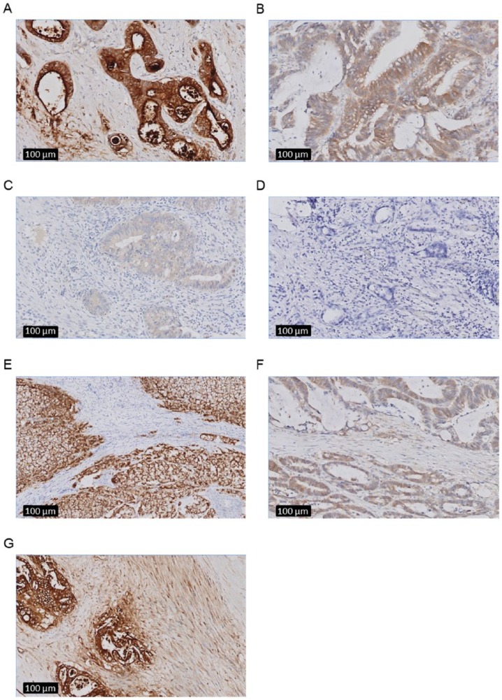
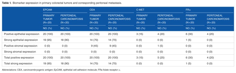
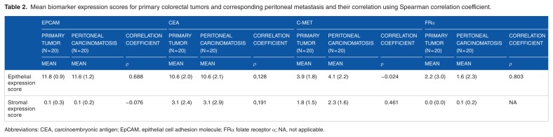
Peritoneal metastasis (PM) occurs in about 10% of patients with colorectal cancer (CRC). Fluorescence imaging can enhance contrast between cancerous and benign tissue, enabling the surgeon to clearly visualize PM during cytoreductive surgery. This study assessed the suitability of different biomarkers as potential targets for tumor-targeted imaging of PM of CRC. Tissue samples from primary tumor and PM from patients with CRC were obtained from the pathology archives and immunohistochemical stainings were performed. Overexpression of the epithelial cell adhesion molecule (EpCAM) and carcinoembryonic antigen (CEA) was seen in 100% of PM samples and the expression was strong in >70% of samples. Tyrosine-kinase Met (C-Met) and folate receptor α overexpression was seen in 20% of PM samples. For successful application of tumor-targeted intraoperative fluorescence imaging of PM, biomarkers need to be identified. We demonstrated that both EpCAM and CEA are suitable targets for fluorescence imaging of PM in patients with CRC.


 分享
分享

 求助内容:
求助内容: 应助结果提醒方式:
 扫码关注我们
