Ji Won Moon, James Ki Shinn, Dalsung Ryu, Se-Yang Oh, Yu Shik Shim, Seung Hwan Yoon
{"title":"骨盆发生率不仅会因年龄和性别而改变,也会因成像时的姿势而改变。","authors":"Ji Won Moon, James Ki Shinn, Dalsung Ryu, Se-Yang Oh, Yu Shik Shim, Seung Hwan Yoon","doi":"10.14245/kjs.2017.14.3.77","DOIUrl":null,"url":null,"abstract":"<p><strong>Objective: </strong>Computed tomography (CT), rather than conventional 2-dimensional radiography, was used to scan and measure pelvic parameters. The results were compared with measurements using X-ray.</p><p><strong>Methods: </strong>Pelvic parameters were measured using both CT and X-ray in 254 patients who underwent both abdomino-pelvic CT and X-ray at the pelvic site. We assessed the similarity of the pelvic parameters between the 2 exams, as well as the correlations of pelvic parameters with sex and age.</p><p><strong>Results: </strong>The mean values of the subjects' pelvic parameters measured on X-ray were: sacral slope (SS), 31.6°; pelvic tilt (PT), 18.6°; and pelvic incidence (PI), 50.2°. The mean values measured on CT were: SS, 35.1°; PT, 11.9°; and PI, 47.0°. PT was found to be 4.07° higher on X-ray and 2.98° higher on CT in women, with these differences being statistically significant (p<0.001, p<0.001). PI was 4.10° higher on X-ray and 2.78° higher on CT in women, with these differences also being statistically significant (p<0.001, p=0.009). We also observed a correlation between age and PI. For men, this correlation coefficient was 0.199 measured using X-ray and 0.184 measured using CT. For women, this correlation coefficient was 0.423 measured using X-ray and 0.372 measured using CT.</p><p><strong>Conclusion: </strong>When measured using CT compared to X-ray, SS increased by 3.5°, PT decreased by 6.7°, and PI decreased by 3.2°. There were also statistically significant differences in PT and PI between male and female subjects, while PI was found to increase with age.</p>","PeriodicalId":17867,"journal":{"name":"Korean Journal of Spine","volume":"14 3","pages":"77-83"},"PeriodicalIF":0.0000,"publicationDate":"2017-09-01","publicationTypes":"Journal Article","fieldsOfStudy":null,"isOpenAccess":false,"openAccessPdf":"https://ftp.ncbi.nlm.nih.gov/pub/pmc/oa_pdf/25/5a/kjs-14-3-77.PMC5642093.pdf","citationCount":"0","resultStr":"{\"title\":\"Pelvic Incidence Can Be Changed not only by Age and Sex, but also by Posture Used during Imaging.\",\"authors\":\"Ji Won Moon, James Ki Shinn, Dalsung Ryu, Se-Yang Oh, Yu Shik Shim, Seung Hwan Yoon\",\"doi\":\"10.14245/kjs.2017.14.3.77\",\"DOIUrl\":null,\"url\":null,\"abstract\":\"<p><strong>Objective: </strong>Computed tomography (CT), rather than conventional 2-dimensional radiography, was used to scan and measure pelvic parameters. The results were compared with measurements using X-ray.</p><p><strong>Methods: </strong>Pelvic parameters were measured using both CT and X-ray in 254 patients who underwent both abdomino-pelvic CT and X-ray at the pelvic site. We assessed the similarity of the pelvic parameters between the 2 exams, as well as the correlations of pelvic parameters with sex and age.</p><p><strong>Results: </strong>The mean values of the subjects' pelvic parameters measured on X-ray were: sacral slope (SS), 31.6°; pelvic tilt (PT), 18.6°; and pelvic incidence (PI), 50.2°. The mean values measured on CT were: SS, 35.1°; PT, 11.9°; and PI, 47.0°. PT was found to be 4.07° higher on X-ray and 2.98° higher on CT in women, with these differences being statistically significant (p<0.001, p<0.001). PI was 4.10° higher on X-ray and 2.78° higher on CT in women, with these differences also being statistically significant (p<0.001, p=0.009). We also observed a correlation between age and PI. For men, this correlation coefficient was 0.199 measured using X-ray and 0.184 measured using CT. For women, this correlation coefficient was 0.423 measured using X-ray and 0.372 measured using CT.</p><p><strong>Conclusion: </strong>When measured using CT compared to X-ray, SS increased by 3.5°, PT decreased by 6.7°, and PI decreased by 3.2°. There were also statistically significant differences in PT and PI between male and female subjects, while PI was found to increase with age.</p>\",\"PeriodicalId\":17867,\"journal\":{\"name\":\"Korean Journal of Spine\",\"volume\":\"14 3\",\"pages\":\"77-83\"},\"PeriodicalIF\":0.0000,\"publicationDate\":\"2017-09-01\",\"publicationTypes\":\"Journal Article\",\"fieldsOfStudy\":null,\"isOpenAccess\":false,\"openAccessPdf\":\"https://ftp.ncbi.nlm.nih.gov/pub/pmc/oa_pdf/25/5a/kjs-14-3-77.PMC5642093.pdf\",\"citationCount\":\"0\",\"resultStr\":null,\"platform\":\"Semanticscholar\",\"paperid\":null,\"PeriodicalName\":\"Korean Journal of Spine\",\"FirstCategoryId\":\"1085\",\"ListUrlMain\":\"https://doi.org/10.14245/kjs.2017.14.3.77\",\"RegionNum\":0,\"RegionCategory\":null,\"ArticlePicture\":[],\"TitleCN\":null,\"AbstractTextCN\":null,\"PMCID\":null,\"EPubDate\":\"2017/9/30 0:00:00\",\"PubModel\":\"Epub\",\"JCR\":\"\",\"JCRName\":\"\",\"Score\":null,\"Total\":0}","platform":"Semanticscholar","paperid":null,"PeriodicalName":"Korean Journal of Spine","FirstCategoryId":"1085","ListUrlMain":"https://doi.org/10.14245/kjs.2017.14.3.77","RegionNum":0,"RegionCategory":null,"ArticlePicture":[],"TitleCN":null,"AbstractTextCN":null,"PMCID":null,"EPubDate":"2017/9/30 0:00:00","PubModel":"Epub","JCR":"","JCRName":"","Score":null,"Total":0}
引用次数: 0
摘要
目的:采用计算机断层扫描(CT)而非传统的二维放射摄影来扫描和测量骨盆参数。方法:对 254 名接受 CT 和 X 光检查的患者进行盆腔参数测量:方法:对 254 名同时接受腹盆腔 CT 和骨盆部位 X 光检查的患者进行了骨盆参数的 CT 和 X 光测量。我们评估了两种检查之间盆腔参数的相似性,以及盆腔参数与性别和年龄的相关性:X光测量的受试者骨盆参数平均值为:骶骨斜度(SS),31.6°;骨盆倾斜度(PT),18.6°;骨盆入射角(PI),50.2°。CT 测量的平均值为SS,35.1°;PT,11.9°;PI,47.0°。发现女性的 X 光片 PT 值比 CT 值高 4.07°,PT 值比 CT 值高 2.98°,这些差异具有统计学意义(p 结论:与 X 光片相比,使用 CT 测量时,SS 上升 3.5°,PT 下降 6.7°,PI 下降 3.2°。男性和女性受试者的 PT 和 PI 差异也有统计学意义,而 PI 则随着年龄的增长而增加。
Pelvic Incidence Can Be Changed not only by Age and Sex, but also by Posture Used during Imaging.
Objective: Computed tomography (CT), rather than conventional 2-dimensional radiography, was used to scan and measure pelvic parameters. The results were compared with measurements using X-ray.
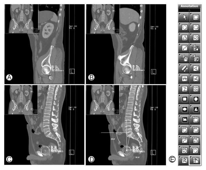
Methods: Pelvic parameters were measured using both CT and X-ray in 254 patients who underwent both abdomino-pelvic CT and X-ray at the pelvic site. We assessed the similarity of the pelvic parameters between the 2 exams, as well as the correlations of pelvic parameters with sex and age.
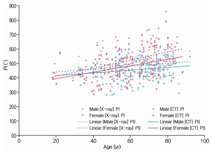
Results: The mean values of the subjects' pelvic parameters measured on X-ray were: sacral slope (SS), 31.6°; pelvic tilt (PT), 18.6°; and pelvic incidence (PI), 50.2°. The mean values measured on CT were: SS, 35.1°; PT, 11.9°; and PI, 47.0°. PT was found to be 4.07° higher on X-ray and 2.98° higher on CT in women, with these differences being statistically significant (p<0.001, p<0.001). PI was 4.10° higher on X-ray and 2.78° higher on CT in women, with these differences also being statistically significant (p<0.001, p=0.009). We also observed a correlation between age and PI. For men, this correlation coefficient was 0.199 measured using X-ray and 0.184 measured using CT. For women, this correlation coefficient was 0.423 measured using X-ray and 0.372 measured using CT.
Conclusion: When measured using CT compared to X-ray, SS increased by 3.5°, PT decreased by 6.7°, and PI decreased by 3.2°. There were also statistically significant differences in PT and PI between male and female subjects, while PI was found to increase with age.


 分享
分享

 求助内容:
求助内容: 应助结果提醒方式:
 扫码关注我们
