{"title":"富血小板血浆双重应用后兔骨钙素和骨连接素的表达。","authors":"Burcu Ozdemir, Bulent Kurtis, Gulay Tuter, Burcu Senguven, Benay Yildirim","doi":"10.17096/jiufd.40536","DOIUrl":null,"url":null,"abstract":"<p><strong>Purpose: </strong>Platelet-rich plasma (PRP) is a novel method for transferring autogenous growth factors to the wound area. The aim of this study was to evaluate the efficacy of double-application of PRP (DA-PRP) on bone healing in rabbit cranial defects by examining osteonectin (ON) and osteocalcin (OC) expression.</p><p><strong>Materials and methods: </strong>Twenty-eight rabbits, each with two surgically prepared calvarial bone defects, were included in this study and divided into six groups: The defects (N=56) were treated with either a single-application of PRP (SA-PRP) (n=10), a combination of SA-PRP and betatricalciumphosphate (SA-PRP+β-TCP) (n=10), only DAPRP (n=8), both DA-PRP and beta-tricalciumphosphate (DA-PRP+β-TCP) (n=8), only beta-tricalciumphosphate (β-TCP) (n=10), or controls (n=10). The animals were sacrificed at 30th day postoperatively and samples were immunohistochemically examined for ON and OC expressions.</p><p><strong>Results: </strong>It was determined that DA-PRP did not significantly improve the ON and OC percentages achieved by SA-PRP or the controls. The three groups treated with β-TCP showed a higher percentage of ON than those treated without β-TCP (p<0.05). The β-TCP treated groups and SA-PRP group demonstrated higher OC percentage than DA-PRP and control groups (p<0.05).</p><p><strong>Conclusion: </strong>The present findings suggest that DAPRP did not have a significant effect on the healing of non-critical size rabbit cranial bone defects.</p>","PeriodicalId":30947,"journal":{"name":"Journal of Istanbul University Faculty of Dentistry","volume":"50 2","pages":"1-9"},"PeriodicalIF":0.0000,"publicationDate":"2016-04-01","publicationTypes":"Journal Article","fieldsOfStudy":null,"isOpenAccess":false,"openAccessPdf":"https://ftp.ncbi.nlm.nih.gov/pub/pmc/oa_pdf/86/85/jiufd-050-001-b.PMC5573525.pdf","citationCount":"7","resultStr":"{\"title\":\"Osteocalcin and osteonectin expression after double application of platelet-rich plasma in rabbits.\",\"authors\":\"Burcu Ozdemir, Bulent Kurtis, Gulay Tuter, Burcu Senguven, Benay Yildirim\",\"doi\":\"10.17096/jiufd.40536\",\"DOIUrl\":null,\"url\":null,\"abstract\":\"<p><strong>Purpose: </strong>Platelet-rich plasma (PRP) is a novel method for transferring autogenous growth factors to the wound area. The aim of this study was to evaluate the efficacy of double-application of PRP (DA-PRP) on bone healing in rabbit cranial defects by examining osteonectin (ON) and osteocalcin (OC) expression.</p><p><strong>Materials and methods: </strong>Twenty-eight rabbits, each with two surgically prepared calvarial bone defects, were included in this study and divided into six groups: The defects (N=56) were treated with either a single-application of PRP (SA-PRP) (n=10), a combination of SA-PRP and betatricalciumphosphate (SA-PRP+β-TCP) (n=10), only DAPRP (n=8), both DA-PRP and beta-tricalciumphosphate (DA-PRP+β-TCP) (n=8), only beta-tricalciumphosphate (β-TCP) (n=10), or controls (n=10). The animals were sacrificed at 30th day postoperatively and samples were immunohistochemically examined for ON and OC expressions.</p><p><strong>Results: </strong>It was determined that DA-PRP did not significantly improve the ON and OC percentages achieved by SA-PRP or the controls. The three groups treated with β-TCP showed a higher percentage of ON than those treated without β-TCP (p<0.05). The β-TCP treated groups and SA-PRP group demonstrated higher OC percentage than DA-PRP and control groups (p<0.05).</p><p><strong>Conclusion: </strong>The present findings suggest that DAPRP did not have a significant effect on the healing of non-critical size rabbit cranial bone defects.</p>\",\"PeriodicalId\":30947,\"journal\":{\"name\":\"Journal of Istanbul University Faculty of Dentistry\",\"volume\":\"50 2\",\"pages\":\"1-9\"},\"PeriodicalIF\":0.0000,\"publicationDate\":\"2016-04-01\",\"publicationTypes\":\"Journal Article\",\"fieldsOfStudy\":null,\"isOpenAccess\":false,\"openAccessPdf\":\"https://ftp.ncbi.nlm.nih.gov/pub/pmc/oa_pdf/86/85/jiufd-050-001-b.PMC5573525.pdf\",\"citationCount\":\"7\",\"resultStr\":null,\"platform\":\"Semanticscholar\",\"paperid\":null,\"PeriodicalName\":\"Journal of Istanbul University Faculty of Dentistry\",\"FirstCategoryId\":\"1085\",\"ListUrlMain\":\"https://doi.org/10.17096/jiufd.40536\",\"RegionNum\":0,\"RegionCategory\":null,\"ArticlePicture\":[],\"TitleCN\":null,\"AbstractTextCN\":null,\"PMCID\":null,\"EPubDate\":\"2016/1/1 0:00:00\",\"PubModel\":\"eCollection\",\"JCR\":\"\",\"JCRName\":\"\",\"Score\":null,\"Total\":0}","platform":"Semanticscholar","paperid":null,"PeriodicalName":"Journal of Istanbul University Faculty of Dentistry","FirstCategoryId":"1085","ListUrlMain":"https://doi.org/10.17096/jiufd.40536","RegionNum":0,"RegionCategory":null,"ArticlePicture":[],"TitleCN":null,"AbstractTextCN":null,"PMCID":null,"EPubDate":"2016/1/1 0:00:00","PubModel":"eCollection","JCR":"","JCRName":"","Score":null,"Total":0}
Osteocalcin and osteonectin expression after double application of platelet-rich plasma in rabbits.
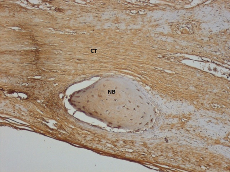
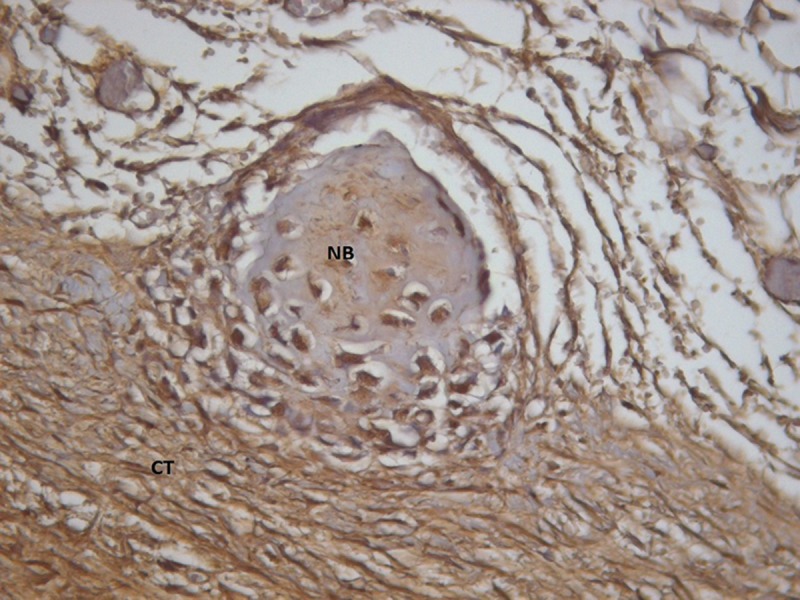
Purpose: Platelet-rich plasma (PRP) is a novel method for transferring autogenous growth factors to the wound area. The aim of this study was to evaluate the efficacy of double-application of PRP (DA-PRP) on bone healing in rabbit cranial defects by examining osteonectin (ON) and osteocalcin (OC) expression.
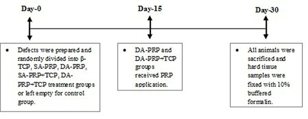
Materials and methods: Twenty-eight rabbits, each with two surgically prepared calvarial bone defects, were included in this study and divided into six groups: The defects (N=56) were treated with either a single-application of PRP (SA-PRP) (n=10), a combination of SA-PRP and betatricalciumphosphate (SA-PRP+β-TCP) (n=10), only DAPRP (n=8), both DA-PRP and beta-tricalciumphosphate (DA-PRP+β-TCP) (n=8), only beta-tricalciumphosphate (β-TCP) (n=10), or controls (n=10). The animals were sacrificed at 30th day postoperatively and samples were immunohistochemically examined for ON and OC expressions.
Results: It was determined that DA-PRP did not significantly improve the ON and OC percentages achieved by SA-PRP or the controls. The three groups treated with β-TCP showed a higher percentage of ON than those treated without β-TCP (p<0.05). The β-TCP treated groups and SA-PRP group demonstrated higher OC percentage than DA-PRP and control groups (p<0.05).
Conclusion: The present findings suggest that DAPRP did not have a significant effect on the healing of non-critical size rabbit cranial bone defects.


 分享
分享

 求助内容:
求助内容: 应助结果提醒方式:
 扫码关注我们
