Meryem Toraman Alkurt, Ilkay Peker, Serife Degerli, Ali Rıza İlker Cebeci, Elif Sadik
{"title":"锥束ct与全景x线检查上颌窦间隔的比较。","authors":"Meryem Toraman Alkurt, Ilkay Peker, Serife Degerli, Ali Rıza İlker Cebeci, Elif Sadik","doi":"10.17096/jiufd.84476","DOIUrl":null,"url":null,"abstract":"<p><strong>Purpose: </strong>The purpose of this retrospective study was to compare the performance of cone-beam computed tomography (CBCT) and panoramic radiography in detecting the presence and location of maxillary sinus septa.</p><p><strong>Materials and methods: </strong>This study included radiographic examination of 104 maxillary sinuses of 52 individuals (26 females, 50% and 26 males, 50%) whose panoramic radiographs and CBCT images were obtained for several dental causes which were examined by the consensus of four dentomaxillofacial radiologists. The posterior maxillary segments in proximity of maxillary sinus were classified as edentulous and dentate maxillary segments. The location of maxillary sinus septa was classified as primary septa and secondary septa according to the presence of maxillary tooth at the affected site. The maxillary sinus septa were divided into three categories (anterior, middle and posterior) according to its relation with posterior maxillary teeth. Data were statistically analyzed with chi-square and Fisher's exact tests.</p><p><strong>Results: </strong>The septa were found in 23.1% and 29.8% of the maxillary sinuses on panoramic radiography and CBCT images, respectively. The majority of maxillary sinus septa were observed in dentate posterior maxillary segments on both panoramic (45.8%) radiography and CBCT (64.5%) images. Statistically significant differences (p<0.001) were found between panoramic radiography and CBCT images for presence, location and neighborhood with the posterior maxillary teeth of maxillary sinus septa.</p><p><strong>Conclusion: </strong>The results of this study demonstrated the low reliability of panoramic radiography images in the detection of maxillary sinus septa. CBCT images can provide valuable information to the clinicians about the presence and location of maxillary sinus septa.</p>","PeriodicalId":30947,"journal":{"name":"Journal of Istanbul University Faculty of Dentistry","volume":"50 3","pages":"8-14"},"PeriodicalIF":0.0000,"publicationDate":"2016-10-01","publicationTypes":"Journal Article","fieldsOfStudy":null,"isOpenAccess":false,"openAccessPdf":"https://sci-hub-pdf.com/10.17096/jiufd.84476","citationCount":"18","resultStr":"{\"title\":\"Comparison of cone-beam computed tomography and panoramic radiographs in detecting maxillary sinus septa.\",\"authors\":\"Meryem Toraman Alkurt, Ilkay Peker, Serife Degerli, Ali Rıza İlker Cebeci, Elif Sadik\",\"doi\":\"10.17096/jiufd.84476\",\"DOIUrl\":null,\"url\":null,\"abstract\":\"<p><strong>Purpose: </strong>The purpose of this retrospective study was to compare the performance of cone-beam computed tomography (CBCT) and panoramic radiography in detecting the presence and location of maxillary sinus septa.</p><p><strong>Materials and methods: </strong>This study included radiographic examination of 104 maxillary sinuses of 52 individuals (26 females, 50% and 26 males, 50%) whose panoramic radiographs and CBCT images were obtained for several dental causes which were examined by the consensus of four dentomaxillofacial radiologists. The posterior maxillary segments in proximity of maxillary sinus were classified as edentulous and dentate maxillary segments. The location of maxillary sinus septa was classified as primary septa and secondary septa according to the presence of maxillary tooth at the affected site. The maxillary sinus septa were divided into three categories (anterior, middle and posterior) according to its relation with posterior maxillary teeth. Data were statistically analyzed with chi-square and Fisher's exact tests.</p><p><strong>Results: </strong>The septa were found in 23.1% and 29.8% of the maxillary sinuses on panoramic radiography and CBCT images, respectively. The majority of maxillary sinus septa were observed in dentate posterior maxillary segments on both panoramic (45.8%) radiography and CBCT (64.5%) images. Statistically significant differences (p<0.001) were found between panoramic radiography and CBCT images for presence, location and neighborhood with the posterior maxillary teeth of maxillary sinus septa.</p><p><strong>Conclusion: </strong>The results of this study demonstrated the low reliability of panoramic radiography images in the detection of maxillary sinus septa. CBCT images can provide valuable information to the clinicians about the presence and location of maxillary sinus septa.</p>\",\"PeriodicalId\":30947,\"journal\":{\"name\":\"Journal of Istanbul University Faculty of Dentistry\",\"volume\":\"50 3\",\"pages\":\"8-14\"},\"PeriodicalIF\":0.0000,\"publicationDate\":\"2016-10-01\",\"publicationTypes\":\"Journal Article\",\"fieldsOfStudy\":null,\"isOpenAccess\":false,\"openAccessPdf\":\"https://sci-hub-pdf.com/10.17096/jiufd.84476\",\"citationCount\":\"18\",\"resultStr\":null,\"platform\":\"Semanticscholar\",\"paperid\":null,\"PeriodicalName\":\"Journal of Istanbul University Faculty of Dentistry\",\"FirstCategoryId\":\"1085\",\"ListUrlMain\":\"https://doi.org/10.17096/jiufd.84476\",\"RegionNum\":0,\"RegionCategory\":null,\"ArticlePicture\":[],\"TitleCN\":null,\"AbstractTextCN\":null,\"PMCID\":null,\"EPubDate\":\"2016/1/1 0:00:00\",\"PubModel\":\"eCollection\",\"JCR\":\"\",\"JCRName\":\"\",\"Score\":null,\"Total\":0}","platform":"Semanticscholar","paperid":null,"PeriodicalName":"Journal of Istanbul University Faculty of Dentistry","FirstCategoryId":"1085","ListUrlMain":"https://doi.org/10.17096/jiufd.84476","RegionNum":0,"RegionCategory":null,"ArticlePicture":[],"TitleCN":null,"AbstractTextCN":null,"PMCID":null,"EPubDate":"2016/1/1 0:00:00","PubModel":"eCollection","JCR":"","JCRName":"","Score":null,"Total":0}
Comparison of cone-beam computed tomography and panoramic radiographs in detecting maxillary sinus septa.
Purpose: The purpose of this retrospective study was to compare the performance of cone-beam computed tomography (CBCT) and panoramic radiography in detecting the presence and location of maxillary sinus septa.
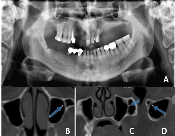
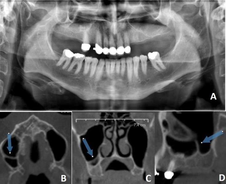
Materials and methods: This study included radiographic examination of 104 maxillary sinuses of 52 individuals (26 females, 50% and 26 males, 50%) whose panoramic radiographs and CBCT images were obtained for several dental causes which were examined by the consensus of four dentomaxillofacial radiologists. The posterior maxillary segments in proximity of maxillary sinus were classified as edentulous and dentate maxillary segments. The location of maxillary sinus septa was classified as primary septa and secondary septa according to the presence of maxillary tooth at the affected site. The maxillary sinus septa were divided into three categories (anterior, middle and posterior) according to its relation with posterior maxillary teeth. Data were statistically analyzed with chi-square and Fisher's exact tests.
Results: The septa were found in 23.1% and 29.8% of the maxillary sinuses on panoramic radiography and CBCT images, respectively. The majority of maxillary sinus septa were observed in dentate posterior maxillary segments on both panoramic (45.8%) radiography and CBCT (64.5%) images. Statistically significant differences (p<0.001) were found between panoramic radiography and CBCT images for presence, location and neighborhood with the posterior maxillary teeth of maxillary sinus septa.
Conclusion: The results of this study demonstrated the low reliability of panoramic radiography images in the detection of maxillary sinus septa. CBCT images can provide valuable information to the clinicians about the presence and location of maxillary sinus septa.


 分享
分享

 求助内容:
求助内容: 应助结果提醒方式:
 扫码关注我们
