Ahmed Almazroa, Sami Alodhayb, Kaamran Raahemifar, Vasudevan Lakshminarayanan
{"title":"青光眼筛查的自动图像处理系统。","authors":"Ahmed Almazroa, Sami Alodhayb, Kaamran Raahemifar, Vasudevan Lakshminarayanan","doi":"10.1155/2017/4826385","DOIUrl":null,"url":null,"abstract":"<p><p>Horizontal and vertical cup to disc ratios are the most crucial parameters used clinically to detect glaucoma or monitor its progress and are manually evaluated from retinal fundus images of the optic nerve head. Due to the rarity of the glaucoma experts as well as the increasing in glaucoma's population, an automatically calculated horizontal and vertical cup to disc ratios (HCDR and VCDR, resp.) can be useful for glaucoma screening. We report on two algorithms to calculate the HCDR and VCDR. In the algorithms, level set and inpainting techniques were developed for segmenting the disc, while thresholding using Type-II fuzzy approach was developed for segmenting the cup. The results from the algorithms were verified using the manual markings of images from a dataset of glaucomatous images (retinal fundus images for glaucoma analysis (RIGA dataset)) by six ophthalmologists. The algorithm's accuracy for HCDR and VCDR combined was 74.2%. Only the accuracy of manual markings by one ophthalmologist was higher than the algorithm's accuracy. The algorithm's best agreement was with markings by ophthalmologist number 1 in 230 images (41.8%) of the total tested images.</p>","PeriodicalId":47063,"journal":{"name":"International Journal of Biomedical Imaging","volume":"2017 ","pages":"4826385"},"PeriodicalIF":1.3000,"publicationDate":"2017-01-01","publicationTypes":"Journal Article","fieldsOfStudy":null,"isOpenAccess":false,"openAccessPdf":"https://sci-hub-pdf.com/10.1155/2017/4826385","citationCount":"26","resultStr":"{\"title\":\"An Automatic Image Processing System for Glaucoma Screening.\",\"authors\":\"Ahmed Almazroa, Sami Alodhayb, Kaamran Raahemifar, Vasudevan Lakshminarayanan\",\"doi\":\"10.1155/2017/4826385\",\"DOIUrl\":null,\"url\":null,\"abstract\":\"<p><p>Horizontal and vertical cup to disc ratios are the most crucial parameters used clinically to detect glaucoma or monitor its progress and are manually evaluated from retinal fundus images of the optic nerve head. Due to the rarity of the glaucoma experts as well as the increasing in glaucoma's population, an automatically calculated horizontal and vertical cup to disc ratios (HCDR and VCDR, resp.) can be useful for glaucoma screening. We report on two algorithms to calculate the HCDR and VCDR. In the algorithms, level set and inpainting techniques were developed for segmenting the disc, while thresholding using Type-II fuzzy approach was developed for segmenting the cup. The results from the algorithms were verified using the manual markings of images from a dataset of glaucomatous images (retinal fundus images for glaucoma analysis (RIGA dataset)) by six ophthalmologists. The algorithm's accuracy for HCDR and VCDR combined was 74.2%. Only the accuracy of manual markings by one ophthalmologist was higher than the algorithm's accuracy. The algorithm's best agreement was with markings by ophthalmologist number 1 in 230 images (41.8%) of the total tested images.</p>\",\"PeriodicalId\":47063,\"journal\":{\"name\":\"International Journal of Biomedical Imaging\",\"volume\":\"2017 \",\"pages\":\"4826385\"},\"PeriodicalIF\":1.3000,\"publicationDate\":\"2017-01-01\",\"publicationTypes\":\"Journal Article\",\"fieldsOfStudy\":null,\"isOpenAccess\":false,\"openAccessPdf\":\"https://sci-hub-pdf.com/10.1155/2017/4826385\",\"citationCount\":\"26\",\"resultStr\":null,\"platform\":\"Semanticscholar\",\"paperid\":null,\"PeriodicalName\":\"International Journal of Biomedical Imaging\",\"FirstCategoryId\":\"1085\",\"ListUrlMain\":\"https://doi.org/10.1155/2017/4826385\",\"RegionNum\":0,\"RegionCategory\":null,\"ArticlePicture\":[],\"TitleCN\":null,\"AbstractTextCN\":null,\"PMCID\":null,\"EPubDate\":\"2017/8/29 0:00:00\",\"PubModel\":\"Epub\",\"JCR\":\"Q2\",\"JCRName\":\"ENGINEERING, BIOMEDICAL\",\"Score\":null,\"Total\":0}","platform":"Semanticscholar","paperid":null,"PeriodicalName":"International Journal of Biomedical Imaging","FirstCategoryId":"1085","ListUrlMain":"https://doi.org/10.1155/2017/4826385","RegionNum":0,"RegionCategory":null,"ArticlePicture":[],"TitleCN":null,"AbstractTextCN":null,"PMCID":null,"EPubDate":"2017/8/29 0:00:00","PubModel":"Epub","JCR":"Q2","JCRName":"ENGINEERING, BIOMEDICAL","Score":null,"Total":0}
An Automatic Image Processing System for Glaucoma Screening.
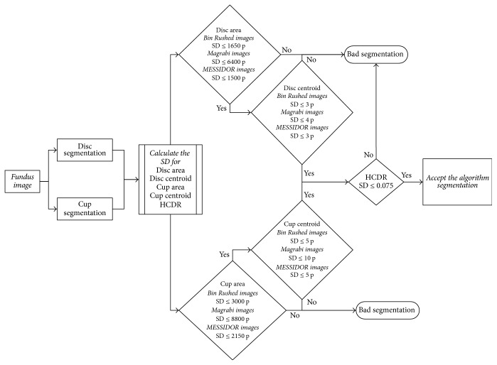
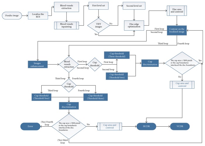
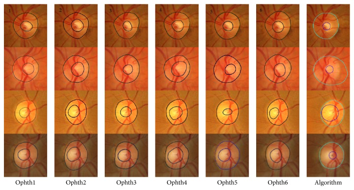
Horizontal and vertical cup to disc ratios are the most crucial parameters used clinically to detect glaucoma or monitor its progress and are manually evaluated from retinal fundus images of the optic nerve head. Due to the rarity of the glaucoma experts as well as the increasing in glaucoma's population, an automatically calculated horizontal and vertical cup to disc ratios (HCDR and VCDR, resp.) can be useful for glaucoma screening. We report on two algorithms to calculate the HCDR and VCDR. In the algorithms, level set and inpainting techniques were developed for segmenting the disc, while thresholding using Type-II fuzzy approach was developed for segmenting the cup. The results from the algorithms were verified using the manual markings of images from a dataset of glaucomatous images (retinal fundus images for glaucoma analysis (RIGA dataset)) by six ophthalmologists. The algorithm's accuracy for HCDR and VCDR combined was 74.2%. Only the accuracy of manual markings by one ophthalmologist was higher than the algorithm's accuracy. The algorithm's best agreement was with markings by ophthalmologist number 1 in 230 images (41.8%) of the total tested images.
期刊介绍:
The International Journal of Biomedical Imaging is managed by a board of editors comprising internationally renowned active researchers. The journal is freely accessible online and also offered for purchase in print format. It employs a web-based review system to ensure swift turnaround times while maintaining high standards. In addition to regular issues, special issues are organized by guest editors. The subject areas covered include (but are not limited to):
Digital radiography and tomosynthesis
X-ray computed tomography (CT)
Magnetic resonance imaging (MRI)
Single photon emission computed tomography (SPECT)
Positron emission tomography (PET)
Ultrasound imaging
Diffuse optical tomography, coherence, fluorescence, bioluminescence tomography, impedance tomography
Neutron imaging for biomedical applications
Magnetic and optical spectroscopy, and optical biopsy
Optical, electron, scanning tunneling/atomic force microscopy
Small animal imaging
Functional, cellular, and molecular imaging
Imaging assays for screening and molecular analysis
Microarray image analysis and bioinformatics
Emerging biomedical imaging techniques
Imaging modality fusion
Biomedical imaging instrumentation
Biomedical image processing, pattern recognition, and analysis
Biomedical image visualization, compression, transmission, and storage
Imaging and modeling related to systems biology and systems biomedicine
Applied mathematics, applied physics, and chemistry related to biomedical imaging
Grid-enabling technology for biomedical imaging and informatics


 分享
分享
 求助内容:
求助内容: 应助结果提醒方式:
 扫码关注我们
