Onur Dincer Kose, Cem Tanyel, Taha Emre Kose, Mehmet Ali Erdem, Abdulkadir Burak Cankaya
{"title":"颌面部海绵状血管瘤患者拔牙1例。","authors":"Onur Dincer Kose, Cem Tanyel, Taha Emre Kose, Mehmet Ali Erdem, Abdulkadir Burak Cankaya","doi":"10.17096/jiufd.77437","DOIUrl":null,"url":null,"abstract":"<p><p>Hemangiomas are benign vascular tumors which represent a rapid growth pattern followed by the involution phase. Generally, they are located in the soft tissues and are usually diagnosed in the first decade of life. Hemangiomas are mostly asymptomatic and rarely affect jaw bones. Mandible is affected more often than maxilla. If there is no complication present, treatment may not be necessary. Treatment planning of hemangiomas should be done by considering the location and the size of the lesion as well as the proximity to vital anatomical structures. The aim of this case report is to describe the procedures of tooth extraction in a patient who had been diagnosed as having maxillary cavernous hemangioma.</p>","PeriodicalId":30947,"journal":{"name":"Journal of Istanbul University Faculty of Dentistry","volume":"50 1","pages":"51-54"},"PeriodicalIF":0.0000,"publicationDate":"2016-01-12","publicationTypes":"Journal Article","fieldsOfStudy":null,"isOpenAccess":false,"openAccessPdf":"https://ftp.ncbi.nlm.nih.gov/pub/pmc/oa_pdf/79/a2/jiufd-050-051.PMC5573453.pdf","citationCount":"0","resultStr":"{\"title\":\"Tooth extraction from a patient with cavernous hemangioma in maxillofacial region: case report.\",\"authors\":\"Onur Dincer Kose, Cem Tanyel, Taha Emre Kose, Mehmet Ali Erdem, Abdulkadir Burak Cankaya\",\"doi\":\"10.17096/jiufd.77437\",\"DOIUrl\":null,\"url\":null,\"abstract\":\"<p><p>Hemangiomas are benign vascular tumors which represent a rapid growth pattern followed by the involution phase. Generally, they are located in the soft tissues and are usually diagnosed in the first decade of life. Hemangiomas are mostly asymptomatic and rarely affect jaw bones. Mandible is affected more often than maxilla. If there is no complication present, treatment may not be necessary. Treatment planning of hemangiomas should be done by considering the location and the size of the lesion as well as the proximity to vital anatomical structures. The aim of this case report is to describe the procedures of tooth extraction in a patient who had been diagnosed as having maxillary cavernous hemangioma.</p>\",\"PeriodicalId\":30947,\"journal\":{\"name\":\"Journal of Istanbul University Faculty of Dentistry\",\"volume\":\"50 1\",\"pages\":\"51-54\"},\"PeriodicalIF\":0.0000,\"publicationDate\":\"2016-01-12\",\"publicationTypes\":\"Journal Article\",\"fieldsOfStudy\":null,\"isOpenAccess\":false,\"openAccessPdf\":\"https://ftp.ncbi.nlm.nih.gov/pub/pmc/oa_pdf/79/a2/jiufd-050-051.PMC5573453.pdf\",\"citationCount\":\"0\",\"resultStr\":null,\"platform\":\"Semanticscholar\",\"paperid\":null,\"PeriodicalName\":\"Journal of Istanbul University Faculty of Dentistry\",\"FirstCategoryId\":\"1085\",\"ListUrlMain\":\"https://doi.org/10.17096/jiufd.77437\",\"RegionNum\":0,\"RegionCategory\":null,\"ArticlePicture\":[],\"TitleCN\":null,\"AbstractTextCN\":null,\"PMCID\":null,\"EPubDate\":\"2016/1/1 0:00:00\",\"PubModel\":\"eCollection\",\"JCR\":\"\",\"JCRName\":\"\",\"Score\":null,\"Total\":0}","platform":"Semanticscholar","paperid":null,"PeriodicalName":"Journal of Istanbul University Faculty of Dentistry","FirstCategoryId":"1085","ListUrlMain":"https://doi.org/10.17096/jiufd.77437","RegionNum":0,"RegionCategory":null,"ArticlePicture":[],"TitleCN":null,"AbstractTextCN":null,"PMCID":null,"EPubDate":"2016/1/1 0:00:00","PubModel":"eCollection","JCR":"","JCRName":"","Score":null,"Total":0}
Tooth extraction from a patient with cavernous hemangioma in maxillofacial region: case report.
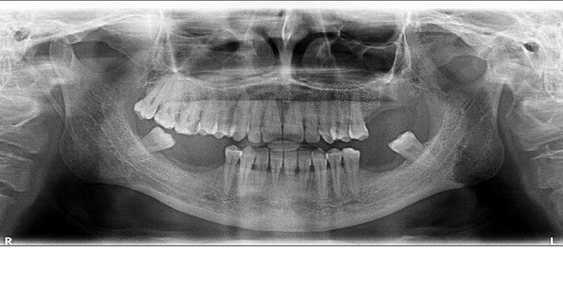
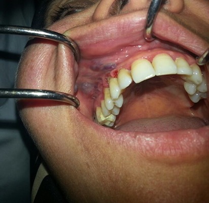
Hemangiomas are benign vascular tumors which represent a rapid growth pattern followed by the involution phase. Generally, they are located in the soft tissues and are usually diagnosed in the first decade of life. Hemangiomas are mostly asymptomatic and rarely affect jaw bones. Mandible is affected more often than maxilla. If there is no complication present, treatment may not be necessary. Treatment planning of hemangiomas should be done by considering the location and the size of the lesion as well as the proximity to vital anatomical structures. The aim of this case report is to describe the procedures of tooth extraction in a patient who had been diagnosed as having maxillary cavernous hemangioma.


 分享
分享

 求助内容:
求助内容: 应助结果提醒方式:
 扫码关注我们
