{"title":"腰2-3段腰椎间盘碎片后硬膜外突出,模拟硬膜外血肿。","authors":"Jin-Sang Kil, Jong-Tae Park","doi":"10.14245/kjs.2017.14.3.115","DOIUrl":null,"url":null,"abstract":"<p><p>Lumbar disk herniation is common. Because of the posterior longitudinal ligament, migration usually occurs into the ventral epidural space. Rarely, fragments migrate into the dorsal epidural space. A 57-year-old man presented with lower back pain and weakness on right hip flexion and right knee flexion. He had lower back pain 1 day previously and received a transforaminal epidural block at a local hospital. The next day, he reported weakness of the right lower extremity. Lumbar spine magnetic resonance imaging revealed a dorsal epidural lesion with compression of the thecal sac at L2-3. Initial differential diagnoses included epidural hematoma after the block, neoplasm, and a sequestrated disk. Posterior lumbar decompression was performed. The lesion was identified intraoperatively as a large herniated disk fragment. Posterior epidural herniation of a lumbar disk fragment is rare and may be difficult to diagnose preoperatively. It may present as a variety of clinical scenarios and, as in this case, may mimic epidural hematoma.</p>","PeriodicalId":17867,"journal":{"name":"Korean Journal of Spine","volume":"14 3","pages":"115-117"},"PeriodicalIF":0.0000,"publicationDate":"2017-09-01","publicationTypes":"Journal Article","fieldsOfStudy":null,"isOpenAccess":false,"openAccessPdf":"https://ftp.ncbi.nlm.nih.gov/pub/pmc/oa_pdf/2b/9a/kjs-14-3-115.PMC5642095.pdf","citationCount":"11","resultStr":"{\"title\":\"Posterior Epidural Herniation of a Lumbar Disk Fragment at L2-3 That Mimicked an Epidural Hematoma.\",\"authors\":\"Jin-Sang Kil, Jong-Tae Park\",\"doi\":\"10.14245/kjs.2017.14.3.115\",\"DOIUrl\":null,\"url\":null,\"abstract\":\"<p><p>Lumbar disk herniation is common. Because of the posterior longitudinal ligament, migration usually occurs into the ventral epidural space. Rarely, fragments migrate into the dorsal epidural space. A 57-year-old man presented with lower back pain and weakness on right hip flexion and right knee flexion. He had lower back pain 1 day previously and received a transforaminal epidural block at a local hospital. The next day, he reported weakness of the right lower extremity. Lumbar spine magnetic resonance imaging revealed a dorsal epidural lesion with compression of the thecal sac at L2-3. Initial differential diagnoses included epidural hematoma after the block, neoplasm, and a sequestrated disk. Posterior lumbar decompression was performed. The lesion was identified intraoperatively as a large herniated disk fragment. Posterior epidural herniation of a lumbar disk fragment is rare and may be difficult to diagnose preoperatively. It may present as a variety of clinical scenarios and, as in this case, may mimic epidural hematoma.</p>\",\"PeriodicalId\":17867,\"journal\":{\"name\":\"Korean Journal of Spine\",\"volume\":\"14 3\",\"pages\":\"115-117\"},\"PeriodicalIF\":0.0000,\"publicationDate\":\"2017-09-01\",\"publicationTypes\":\"Journal Article\",\"fieldsOfStudy\":null,\"isOpenAccess\":false,\"openAccessPdf\":\"https://ftp.ncbi.nlm.nih.gov/pub/pmc/oa_pdf/2b/9a/kjs-14-3-115.PMC5642095.pdf\",\"citationCount\":\"11\",\"resultStr\":null,\"platform\":\"Semanticscholar\",\"paperid\":null,\"PeriodicalName\":\"Korean Journal of Spine\",\"FirstCategoryId\":\"1085\",\"ListUrlMain\":\"https://doi.org/10.14245/kjs.2017.14.3.115\",\"RegionNum\":0,\"RegionCategory\":null,\"ArticlePicture\":[],\"TitleCN\":null,\"AbstractTextCN\":null,\"PMCID\":null,\"EPubDate\":\"2017/9/30 0:00:00\",\"PubModel\":\"Epub\",\"JCR\":\"\",\"JCRName\":\"\",\"Score\":null,\"Total\":0}","platform":"Semanticscholar","paperid":null,"PeriodicalName":"Korean Journal of Spine","FirstCategoryId":"1085","ListUrlMain":"https://doi.org/10.14245/kjs.2017.14.3.115","RegionNum":0,"RegionCategory":null,"ArticlePicture":[],"TitleCN":null,"AbstractTextCN":null,"PMCID":null,"EPubDate":"2017/9/30 0:00:00","PubModel":"Epub","JCR":"","JCRName":"","Score":null,"Total":0}
Posterior Epidural Herniation of a Lumbar Disk Fragment at L2-3 That Mimicked an Epidural Hematoma.
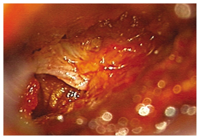
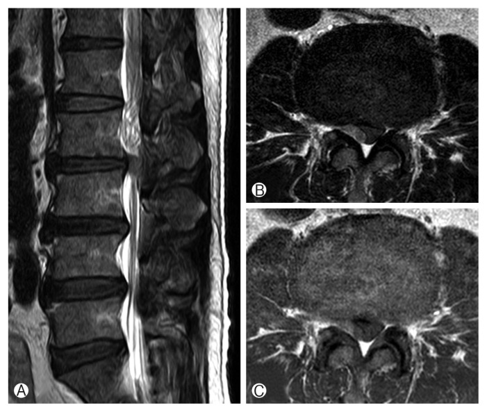
Lumbar disk herniation is common. Because of the posterior longitudinal ligament, migration usually occurs into the ventral epidural space. Rarely, fragments migrate into the dorsal epidural space. A 57-year-old man presented with lower back pain and weakness on right hip flexion and right knee flexion. He had lower back pain 1 day previously and received a transforaminal epidural block at a local hospital. The next day, he reported weakness of the right lower extremity. Lumbar spine magnetic resonance imaging revealed a dorsal epidural lesion with compression of the thecal sac at L2-3. Initial differential diagnoses included epidural hematoma after the block, neoplasm, and a sequestrated disk. Posterior lumbar decompression was performed. The lesion was identified intraoperatively as a large herniated disk fragment. Posterior epidural herniation of a lumbar disk fragment is rare and may be difficult to diagnose preoperatively. It may present as a variety of clinical scenarios and, as in this case, may mimic epidural hematoma.


 分享
分享

 求助内容:
求助内容: 应助结果提醒方式:
 扫码关注我们
