Burak Unlu, Ziya Ayhan, Banu Lebe, Suleyman Men, Ismet Durak, Ali Osman Saatci
{"title":"单侧成人黄色肉芽肿性结膜、角膜缘和巩膜浸润导致眼动脉和视网膜中央静脉联合闭塞。","authors":"Burak Unlu, Ziya Ayhan, Banu Lebe, Suleyman Men, Ismet Durak, Ali Osman Saatci","doi":"10.2174/1874364101711010362","DOIUrl":null,"url":null,"abstract":"<p><strong>Objective: </strong>To describe the features of a female patient with a biopsy-proven xanthogranulomatous infiltration of the conjunctiva, limbus and sclera who had an exudative retinal detachment, combined ophthalmic artery and central retinal vein occlusion unilaterally.</p><p><strong>Method: </strong>A-53-year old otherwise healthy woman presenting with a painful visual loss in her right eye underwent an ophthalmic examination, meticulous systemic work-up and histopathologic assessment.</p><p><strong>Results: </strong>Ophthalmic examination revealed multiple subconjunctival masses, upper limbal infiltrations, trace cells in the anterior chamber, pale looking posterior fundus, 360 degree scattered retinal hemorrhages and marked exudative retinal detachment in her right eye. Left eye was completely normal.A biopsy taken from one of the subconjunctival masses demonstrated a diffuse infiltration of the histiocytes and this was interpreted as a xanthogranulomatous infiltration with the help of immunohistochemical staining techniques.</p><p><strong>Conclusion: </strong>Present case is the only reported adult case with xanthogranulomatous-like infiltration of the eyeball featuring both anterior and posterior segment involvement without any concomitant major systemic disturbances.</p>","PeriodicalId":512318,"journal":{"name":"The Open Ophthalmology Journal","volume":"11 ","pages":"362-367"},"PeriodicalIF":0.0000,"publicationDate":"2017-11-23","publicationTypes":"Journal Article","fieldsOfStudy":null,"isOpenAccess":false,"openAccessPdf":"https://www.ncbi.nlm.nih.gov/pmc/articles/PMC5725586/pdf/","citationCount":"0","resultStr":"{\"title\":\"Unilateral Adult Xanthogranulomatous Infiltration of the Conjunctiva, Limbus and Sclera Leading to a Combined Ophthalmic Artery and Central Retinal Vein Occlusion.\",\"authors\":\"Burak Unlu, Ziya Ayhan, Banu Lebe, Suleyman Men, Ismet Durak, Ali Osman Saatci\",\"doi\":\"10.2174/1874364101711010362\",\"DOIUrl\":null,\"url\":null,\"abstract\":\"<p><strong>Objective: </strong>To describe the features of a female patient with a biopsy-proven xanthogranulomatous infiltration of the conjunctiva, limbus and sclera who had an exudative retinal detachment, combined ophthalmic artery and central retinal vein occlusion unilaterally.</p><p><strong>Method: </strong>A-53-year old otherwise healthy woman presenting with a painful visual loss in her right eye underwent an ophthalmic examination, meticulous systemic work-up and histopathologic assessment.</p><p><strong>Results: </strong>Ophthalmic examination revealed multiple subconjunctival masses, upper limbal infiltrations, trace cells in the anterior chamber, pale looking posterior fundus, 360 degree scattered retinal hemorrhages and marked exudative retinal detachment in her right eye. Left eye was completely normal.A biopsy taken from one of the subconjunctival masses demonstrated a diffuse infiltration of the histiocytes and this was interpreted as a xanthogranulomatous infiltration with the help of immunohistochemical staining techniques.</p><p><strong>Conclusion: </strong>Present case is the only reported adult case with xanthogranulomatous-like infiltration of the eyeball featuring both anterior and posterior segment involvement without any concomitant major systemic disturbances.</p>\",\"PeriodicalId\":512318,\"journal\":{\"name\":\"The Open Ophthalmology Journal\",\"volume\":\"11 \",\"pages\":\"362-367\"},\"PeriodicalIF\":0.0000,\"publicationDate\":\"2017-11-23\",\"publicationTypes\":\"Journal Article\",\"fieldsOfStudy\":null,\"isOpenAccess\":false,\"openAccessPdf\":\"https://www.ncbi.nlm.nih.gov/pmc/articles/PMC5725586/pdf/\",\"citationCount\":\"0\",\"resultStr\":null,\"platform\":\"Semanticscholar\",\"paperid\":null,\"PeriodicalName\":\"The Open Ophthalmology Journal\",\"FirstCategoryId\":\"1085\",\"ListUrlMain\":\"https://doi.org/10.2174/1874364101711010362\",\"RegionNum\":0,\"RegionCategory\":null,\"ArticlePicture\":[],\"TitleCN\":null,\"AbstractTextCN\":null,\"PMCID\":null,\"EPubDate\":\"2017/1/1 0:00:00\",\"PubModel\":\"eCollection\",\"JCR\":\"\",\"JCRName\":\"\",\"Score\":null,\"Total\":0}","platform":"Semanticscholar","paperid":null,"PeriodicalName":"The Open Ophthalmology Journal","FirstCategoryId":"1085","ListUrlMain":"https://doi.org/10.2174/1874364101711010362","RegionNum":0,"RegionCategory":null,"ArticlePicture":[],"TitleCN":null,"AbstractTextCN":null,"PMCID":null,"EPubDate":"2017/1/1 0:00:00","PubModel":"eCollection","JCR":"","JCRName":"","Score":null,"Total":0}
Unilateral Adult Xanthogranulomatous Infiltration of the Conjunctiva, Limbus and Sclera Leading to a Combined Ophthalmic Artery and Central Retinal Vein Occlusion.
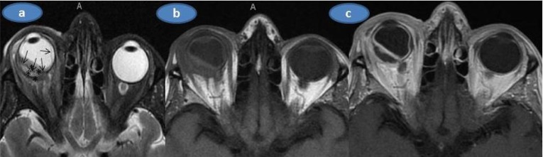
Objective: To describe the features of a female patient with a biopsy-proven xanthogranulomatous infiltration of the conjunctiva, limbus and sclera who had an exudative retinal detachment, combined ophthalmic artery and central retinal vein occlusion unilaterally.
Method: A-53-year old otherwise healthy woman presenting with a painful visual loss in her right eye underwent an ophthalmic examination, meticulous systemic work-up and histopathologic assessment.
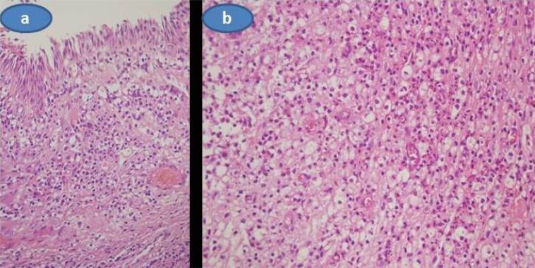
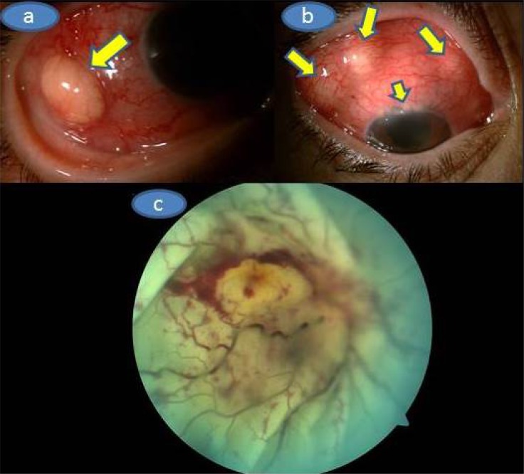
Results: Ophthalmic examination revealed multiple subconjunctival masses, upper limbal infiltrations, trace cells in the anterior chamber, pale looking posterior fundus, 360 degree scattered retinal hemorrhages and marked exudative retinal detachment in her right eye. Left eye was completely normal.A biopsy taken from one of the subconjunctival masses demonstrated a diffuse infiltration of the histiocytes and this was interpreted as a xanthogranulomatous infiltration with the help of immunohistochemical staining techniques.
Conclusion: Present case is the only reported adult case with xanthogranulomatous-like infiltration of the eyeball featuring both anterior and posterior segment involvement without any concomitant major systemic disturbances.


 分享
分享

 求助内容:
求助内容: 应助结果提醒方式:
 扫码关注我们
