{"title":"基于熵的视网膜图像视盘自动定位算法。","authors":"Lamia AbedNoor Muhammed","doi":"10.1155/2018/2815163","DOIUrl":null,"url":null,"abstract":"<p><p>Examining retinal image continuously plays an important role in determining human eye health; with any variation present in this image, it may be resulting from some disease. Therefore, there is a need for computer-aided scanning for retinal image to perform this task automatically and accurately. The fundamental step in this task is identification of the retina elements; optical disk localization is the most important one in this identification. Different optical disc localization algorithms have been suggested, such as an algorithm that would be proposed in this paper. The assumption is based on the fact that optical disc area has rich information, so its entropy value is more significant in this area. The suggested algorithm has recursive steps for testing the entropy of different patches in image; sliding window technique is used to get these patches in a specific way. The results of practical work were obtained using different common data set, which achieved good accuracy in trivial computation time. Finally, this paper consists of four sections: a section for introduction containing the related works, a section for methodology and material, a section for practical work with results, and a section for conclusion.</p>","PeriodicalId":47063,"journal":{"name":"International Journal of Biomedical Imaging","volume":"2018 ","pages":"2815163"},"PeriodicalIF":1.3000,"publicationDate":"2018-02-06","publicationTypes":"Journal Article","fieldsOfStudy":null,"isOpenAccess":false,"openAccessPdf":"https://sci-hub-pdf.com/10.1155/2018/2815163","citationCount":"18","resultStr":"{\"title\":\"Localizing Optic Disc in Retinal Image Automatically with Entropy Based Algorithm.\",\"authors\":\"Lamia AbedNoor Muhammed\",\"doi\":\"10.1155/2018/2815163\",\"DOIUrl\":null,\"url\":null,\"abstract\":\"<p><p>Examining retinal image continuously plays an important role in determining human eye health; with any variation present in this image, it may be resulting from some disease. Therefore, there is a need for computer-aided scanning for retinal image to perform this task automatically and accurately. The fundamental step in this task is identification of the retina elements; optical disk localization is the most important one in this identification. Different optical disc localization algorithms have been suggested, such as an algorithm that would be proposed in this paper. The assumption is based on the fact that optical disc area has rich information, so its entropy value is more significant in this area. The suggested algorithm has recursive steps for testing the entropy of different patches in image; sliding window technique is used to get these patches in a specific way. The results of practical work were obtained using different common data set, which achieved good accuracy in trivial computation time. Finally, this paper consists of four sections: a section for introduction containing the related works, a section for methodology and material, a section for practical work with results, and a section for conclusion.</p>\",\"PeriodicalId\":47063,\"journal\":{\"name\":\"International Journal of Biomedical Imaging\",\"volume\":\"2018 \",\"pages\":\"2815163\"},\"PeriodicalIF\":1.3000,\"publicationDate\":\"2018-02-06\",\"publicationTypes\":\"Journal Article\",\"fieldsOfStudy\":null,\"isOpenAccess\":false,\"openAccessPdf\":\"https://sci-hub-pdf.com/10.1155/2018/2815163\",\"citationCount\":\"18\",\"resultStr\":null,\"platform\":\"Semanticscholar\",\"paperid\":null,\"PeriodicalName\":\"International Journal of Biomedical Imaging\",\"FirstCategoryId\":\"1085\",\"ListUrlMain\":\"https://doi.org/10.1155/2018/2815163\",\"RegionNum\":0,\"RegionCategory\":null,\"ArticlePicture\":[],\"TitleCN\":null,\"AbstractTextCN\":null,\"PMCID\":null,\"EPubDate\":\"2018/1/1 0:00:00\",\"PubModel\":\"eCollection\",\"JCR\":\"Q2\",\"JCRName\":\"ENGINEERING, BIOMEDICAL\",\"Score\":null,\"Total\":0}","platform":"Semanticscholar","paperid":null,"PeriodicalName":"International Journal of Biomedical Imaging","FirstCategoryId":"1085","ListUrlMain":"https://doi.org/10.1155/2018/2815163","RegionNum":0,"RegionCategory":null,"ArticlePicture":[],"TitleCN":null,"AbstractTextCN":null,"PMCID":null,"EPubDate":"2018/1/1 0:00:00","PubModel":"eCollection","JCR":"Q2","JCRName":"ENGINEERING, BIOMEDICAL","Score":null,"Total":0}
Localizing Optic Disc in Retinal Image Automatically with Entropy Based Algorithm.
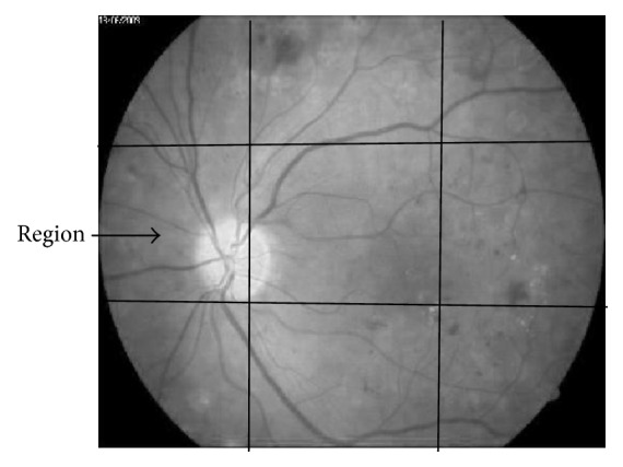
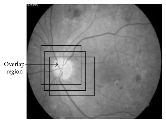
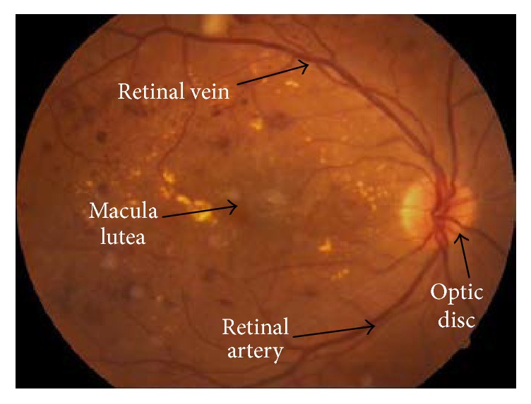
Examining retinal image continuously plays an important role in determining human eye health; with any variation present in this image, it may be resulting from some disease. Therefore, there is a need for computer-aided scanning for retinal image to perform this task automatically and accurately. The fundamental step in this task is identification of the retina elements; optical disk localization is the most important one in this identification. Different optical disc localization algorithms have been suggested, such as an algorithm that would be proposed in this paper. The assumption is based on the fact that optical disc area has rich information, so its entropy value is more significant in this area. The suggested algorithm has recursive steps for testing the entropy of different patches in image; sliding window technique is used to get these patches in a specific way. The results of practical work were obtained using different common data set, which achieved good accuracy in trivial computation time. Finally, this paper consists of four sections: a section for introduction containing the related works, a section for methodology and material, a section for practical work with results, and a section for conclusion.
期刊介绍:
The International Journal of Biomedical Imaging is managed by a board of editors comprising internationally renowned active researchers. The journal is freely accessible online and also offered for purchase in print format. It employs a web-based review system to ensure swift turnaround times while maintaining high standards. In addition to regular issues, special issues are organized by guest editors. The subject areas covered include (but are not limited to):
Digital radiography and tomosynthesis
X-ray computed tomography (CT)
Magnetic resonance imaging (MRI)
Single photon emission computed tomography (SPECT)
Positron emission tomography (PET)
Ultrasound imaging
Diffuse optical tomography, coherence, fluorescence, bioluminescence tomography, impedance tomography
Neutron imaging for biomedical applications
Magnetic and optical spectroscopy, and optical biopsy
Optical, electron, scanning tunneling/atomic force microscopy
Small animal imaging
Functional, cellular, and molecular imaging
Imaging assays for screening and molecular analysis
Microarray image analysis and bioinformatics
Emerging biomedical imaging techniques
Imaging modality fusion
Biomedical imaging instrumentation
Biomedical image processing, pattern recognition, and analysis
Biomedical image visualization, compression, transmission, and storage
Imaging and modeling related to systems biology and systems biomedicine
Applied mathematics, applied physics, and chemistry related to biomedical imaging
Grid-enabling technology for biomedical imaging and informatics


 分享
分享
 求助内容:
求助内容: 应助结果提醒方式:
 扫码关注我们
