William D Dunn, Hugo J W L Aerts, Lee A Cooper, Chad A Holder, Scott N Hwang, Carle C Jaffe, Daniel J Brat, Rajan Jain, Adam E Flanders, Pascal O Zinn, Rivka R Colen, David A Gutman
{"title":"评估软件平台对胶质母细胞瘤体积分割的影响","authors":"William D Dunn, Hugo J W L Aerts, Lee A Cooper, Chad A Holder, Scott N Hwang, Carle C Jaffe, Daniel J Brat, Rajan Jain, Adam E Flanders, Pascal O Zinn, Rivka R Colen, David A Gutman","doi":"10.17756/jnpn.2016-008","DOIUrl":null,"url":null,"abstract":"<p><strong>Background: </strong>Radiological assessments of biologically relevant regions in glioblastoma have been associated with genotypic characteristics, implying a potential role in personalized medicine. Here, we assess the reproducibility and association with survival of two volumetric segmentation platforms and explore how methodology could impact subsequent interpretation and analysis.</p><p><strong>Methods: </strong>Post-contrast T1- and T2-weighted FLAIR MR images of 67 TCGA patients were segmented into five distinct compartments (necrosis, contrast-enhancement, FLAIR, post contrast abnormal, and total abnormal tumor volumes) by two quantitative image segmentation platforms - 3D Slicer and a method based on Velocity AI and FSL. We investigated the internal consistency of each platform by correlation statistics, association with survival, and concordance with consensus neuroradiologist ratings using ordinal logistic regression.</p><p><strong>Results: </strong>We found high correlations between the two platforms for FLAIR, post contrast abnormal, and total abnormal tumor volumes (spearman's r(67) = 0.952, 0.959, and 0.969 respectively). Only modest agreement was observed for necrosis and contrast-enhancement volumes (r(67) = 0.693 and 0.773 respectively), likely arising from differences in manual and automated segmentation methods of these regions by 3D Slicer and Velocity AI/FSL, respectively. Survival analysis based on AUC revealed significant predictive power of both platforms for the following volumes: contrast-enhancement, post contrast abnormal, and total abnormal tumor volumes. Finally, ordinal logistic regression demonstrated correspondence to manual ratings for several features.</p><p><strong>Conclusion: </strong>Tumor volume measurements from both volumetric platforms produced highly concordant and reproducible estimates across platforms for general features. As automated or semi-automated volumetric measurements replace manual linear or area measurements, it will become increasingly important to keep in mind that measurement differences between segmentation platforms for more detailed features could influence downstream survival or radio genomic analyses.</p>","PeriodicalId":91910,"journal":{"name":"Journal of neuroimaging in psychiatry & neurology","volume":"1 2","pages":"64-72"},"PeriodicalIF":0.0000,"publicationDate":"2016-01-01","publicationTypes":"Journal Article","fieldsOfStudy":null,"isOpenAccess":false,"openAccessPdf":"https://www.ncbi.nlm.nih.gov/pmc/articles/PMC5870135/pdf/","citationCount":"0","resultStr":"{\"title\":\"Assessing the Effects of Software Platforms on Volumetric Segmentation of Glioblastoma.\",\"authors\":\"William D Dunn, Hugo J W L Aerts, Lee A Cooper, Chad A Holder, Scott N Hwang, Carle C Jaffe, Daniel J Brat, Rajan Jain, Adam E Flanders, Pascal O Zinn, Rivka R Colen, David A Gutman\",\"doi\":\"10.17756/jnpn.2016-008\",\"DOIUrl\":null,\"url\":null,\"abstract\":\"<p><strong>Background: </strong>Radiological assessments of biologically relevant regions in glioblastoma have been associated with genotypic characteristics, implying a potential role in personalized medicine. Here, we assess the reproducibility and association with survival of two volumetric segmentation platforms and explore how methodology could impact subsequent interpretation and analysis.</p><p><strong>Methods: </strong>Post-contrast T1- and T2-weighted FLAIR MR images of 67 TCGA patients were segmented into five distinct compartments (necrosis, contrast-enhancement, FLAIR, post contrast abnormal, and total abnormal tumor volumes) by two quantitative image segmentation platforms - 3D Slicer and a method based on Velocity AI and FSL. We investigated the internal consistency of each platform by correlation statistics, association with survival, and concordance with consensus neuroradiologist ratings using ordinal logistic regression.</p><p><strong>Results: </strong>We found high correlations between the two platforms for FLAIR, post contrast abnormal, and total abnormal tumor volumes (spearman's r(67) = 0.952, 0.959, and 0.969 respectively). Only modest agreement was observed for necrosis and contrast-enhancement volumes (r(67) = 0.693 and 0.773 respectively), likely arising from differences in manual and automated segmentation methods of these regions by 3D Slicer and Velocity AI/FSL, respectively. Survival analysis based on AUC revealed significant predictive power of both platforms for the following volumes: contrast-enhancement, post contrast abnormal, and total abnormal tumor volumes. Finally, ordinal logistic regression demonstrated correspondence to manual ratings for several features.</p><p><strong>Conclusion: </strong>Tumor volume measurements from both volumetric platforms produced highly concordant and reproducible estimates across platforms for general features. As automated or semi-automated volumetric measurements replace manual linear or area measurements, it will become increasingly important to keep in mind that measurement differences between segmentation platforms for more detailed features could influence downstream survival or radio genomic analyses.</p>\",\"PeriodicalId\":91910,\"journal\":{\"name\":\"Journal of neuroimaging in psychiatry & neurology\",\"volume\":\"1 2\",\"pages\":\"64-72\"},\"PeriodicalIF\":0.0000,\"publicationDate\":\"2016-01-01\",\"publicationTypes\":\"Journal Article\",\"fieldsOfStudy\":null,\"isOpenAccess\":false,\"openAccessPdf\":\"https://www.ncbi.nlm.nih.gov/pmc/articles/PMC5870135/pdf/\",\"citationCount\":\"0\",\"resultStr\":null,\"platform\":\"Semanticscholar\",\"paperid\":null,\"PeriodicalName\":\"Journal of neuroimaging in psychiatry & neurology\",\"FirstCategoryId\":\"1085\",\"ListUrlMain\":\"https://doi.org/10.17756/jnpn.2016-008\",\"RegionNum\":0,\"RegionCategory\":null,\"ArticlePicture\":[],\"TitleCN\":null,\"AbstractTextCN\":null,\"PMCID\":null,\"EPubDate\":\"2016/7/20 0:00:00\",\"PubModel\":\"Epub\",\"JCR\":\"\",\"JCRName\":\"\",\"Score\":null,\"Total\":0}","platform":"Semanticscholar","paperid":null,"PeriodicalName":"Journal of neuroimaging in psychiatry & neurology","FirstCategoryId":"1085","ListUrlMain":"https://doi.org/10.17756/jnpn.2016-008","RegionNum":0,"RegionCategory":null,"ArticlePicture":[],"TitleCN":null,"AbstractTextCN":null,"PMCID":null,"EPubDate":"2016/7/20 0:00:00","PubModel":"Epub","JCR":"","JCRName":"","Score":null,"Total":0}
Assessing the Effects of Software Platforms on Volumetric Segmentation of Glioblastoma.
Background: Radiological assessments of biologically relevant regions in glioblastoma have been associated with genotypic characteristics, implying a potential role in personalized medicine. Here, we assess the reproducibility and association with survival of two volumetric segmentation platforms and explore how methodology could impact subsequent interpretation and analysis.
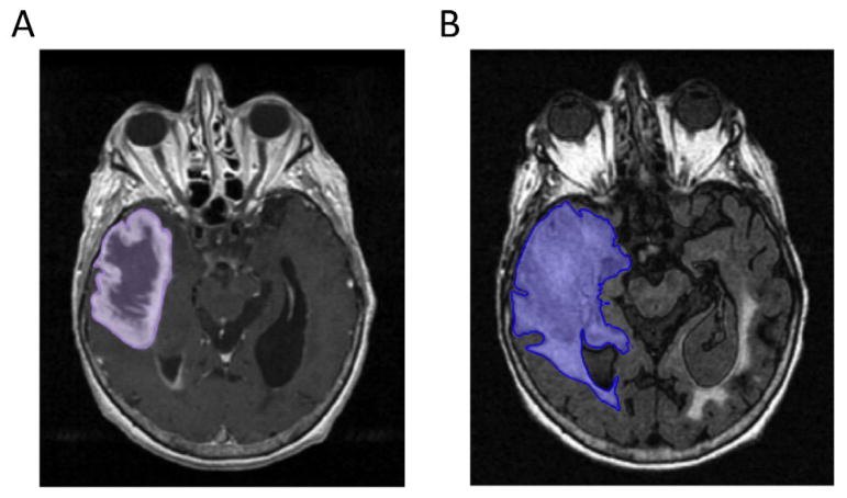
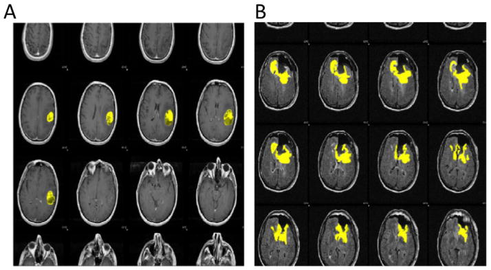
Methods: Post-contrast T1- and T2-weighted FLAIR MR images of 67 TCGA patients were segmented into five distinct compartments (necrosis, contrast-enhancement, FLAIR, post contrast abnormal, and total abnormal tumor volumes) by two quantitative image segmentation platforms - 3D Slicer and a method based on Velocity AI and FSL. We investigated the internal consistency of each platform by correlation statistics, association with survival, and concordance with consensus neuroradiologist ratings using ordinal logistic regression.
Results: We found high correlations between the two platforms for FLAIR, post contrast abnormal, and total abnormal tumor volumes (spearman's r(67) = 0.952, 0.959, and 0.969 respectively). Only modest agreement was observed for necrosis and contrast-enhancement volumes (r(67) = 0.693 and 0.773 respectively), likely arising from differences in manual and automated segmentation methods of these regions by 3D Slicer and Velocity AI/FSL, respectively. Survival analysis based on AUC revealed significant predictive power of both platforms for the following volumes: contrast-enhancement, post contrast abnormal, and total abnormal tumor volumes. Finally, ordinal logistic regression demonstrated correspondence to manual ratings for several features.
Conclusion: Tumor volume measurements from both volumetric platforms produced highly concordant and reproducible estimates across platforms for general features. As automated or semi-automated volumetric measurements replace manual linear or area measurements, it will become increasingly important to keep in mind that measurement differences between segmentation platforms for more detailed features could influence downstream survival or radio genomic analyses.


 分享
分享

 求助内容:
求助内容: 应助结果提醒方式:
 扫码关注我们
