Caroline Ewertsen, Jonathan Carlsen, Mohammed Aftab Perveez, Henrik Schytz
{"title":"健康人颈肩肌肉剪切波弹性成像的参考值。","authors":"Caroline Ewertsen, Jonathan Carlsen, Mohammed Aftab Perveez, Henrik Schytz","doi":"10.1055/s-0044-102013","DOIUrl":null,"url":null,"abstract":"<p><strong>Purpose: </strong>to establish reference values for ultrasound shear-wave elastography for pericranial muscles in healthy individuals (m. trapezius, m. splenius capitis, m. semispinalis capitis, m. sternocleidomastoideus and m. masseter). Also to evaluate day-to-day variations in the shear-wave speeds and evaluate the effect of the pennation of the muscle fibers, ie scanning parallel or perpendicularly to the fibers.</p><p><strong>Materials and methods: </strong>10 healthy individuals (5 males and 5 females) had their pericranial muscles examined with shear-wave elastography in two orthogonal planes on two different days for their dominant and non-dominant side. Mean shear wave speeds from 5 ROI's in each muscle, for each scan plane for the dominant and non-dominant side for the two days were calculated. The effect of the different parameters - muscle pennation, gender, dominant vs non-dominant side and day was evaluated.</p><p><strong>Results: </strong>The effect of scan plane in relation to muscle pennation was statistically significant (p<0.0001). The mean shear-wave speed when scanning parallel to the muscle fibers was significantly higher than the mean shear-wave speed when scanning perpendicularly to the fibers. The day-to-day variation was statistically significant (p=0.0258), but not clinically relevant. Shear-wave speeds differed significantly between muscles. Mean shear wave speeds (m/s) for the muscles in the parallel plane were: for masseter 2.45 (SD:+/-0.25), semispinal 3.36 (SD:+/-0.75), splenius 3.04 (SD:+/-0.65), sternocleidomastoid 2.75 (SD:+/-0.23), trapezius 3.20 (SD:+/-0.27) and trapezius lateral 3.87 (SD:+/-3.87).</p><p><strong>Conclusion: </strong>The shear wave speed variation depended on the direction of scanning. Shear wave elastography may be a method to evaluate muscle stiffness in patients suffering from chronic neck pain.</p>","PeriodicalId":44852,"journal":{"name":"Ultrasound International Open","volume":"4 1","pages":"E23-E29"},"PeriodicalIF":1.6000,"publicationDate":"2018-01-01","publicationTypes":"Journal Article","fieldsOfStudy":null,"isOpenAccess":false,"openAccessPdf":"https://sci-hub-pdf.com/10.1055/s-0044-102013","citationCount":"26","resultStr":"{\"title\":\"Reference Values for Shear Wave Elastography of Neck and Shoulder Muscles in Healthy Individuals.\",\"authors\":\"Caroline Ewertsen, Jonathan Carlsen, Mohammed Aftab Perveez, Henrik Schytz\",\"doi\":\"10.1055/s-0044-102013\",\"DOIUrl\":null,\"url\":null,\"abstract\":\"<p><strong>Purpose: </strong>to establish reference values for ultrasound shear-wave elastography for pericranial muscles in healthy individuals (m. trapezius, m. splenius capitis, m. semispinalis capitis, m. sternocleidomastoideus and m. masseter). Also to evaluate day-to-day variations in the shear-wave speeds and evaluate the effect of the pennation of the muscle fibers, ie scanning parallel or perpendicularly to the fibers.</p><p><strong>Materials and methods: </strong>10 healthy individuals (5 males and 5 females) had their pericranial muscles examined with shear-wave elastography in two orthogonal planes on two different days for their dominant and non-dominant side. Mean shear wave speeds from 5 ROI's in each muscle, for each scan plane for the dominant and non-dominant side for the two days were calculated. The effect of the different parameters - muscle pennation, gender, dominant vs non-dominant side and day was evaluated.</p><p><strong>Results: </strong>The effect of scan plane in relation to muscle pennation was statistically significant (p<0.0001). The mean shear-wave speed when scanning parallel to the muscle fibers was significantly higher than the mean shear-wave speed when scanning perpendicularly to the fibers. The day-to-day variation was statistically significant (p=0.0258), but not clinically relevant. Shear-wave speeds differed significantly between muscles. Mean shear wave speeds (m/s) for the muscles in the parallel plane were: for masseter 2.45 (SD:+/-0.25), semispinal 3.36 (SD:+/-0.75), splenius 3.04 (SD:+/-0.65), sternocleidomastoid 2.75 (SD:+/-0.23), trapezius 3.20 (SD:+/-0.27) and trapezius lateral 3.87 (SD:+/-3.87).</p><p><strong>Conclusion: </strong>The shear wave speed variation depended on the direction of scanning. Shear wave elastography may be a method to evaluate muscle stiffness in patients suffering from chronic neck pain.</p>\",\"PeriodicalId\":44852,\"journal\":{\"name\":\"Ultrasound International Open\",\"volume\":\"4 1\",\"pages\":\"E23-E29\"},\"PeriodicalIF\":1.6000,\"publicationDate\":\"2018-01-01\",\"publicationTypes\":\"Journal Article\",\"fieldsOfStudy\":null,\"isOpenAccess\":false,\"openAccessPdf\":\"https://sci-hub-pdf.com/10.1055/s-0044-102013\",\"citationCount\":\"26\",\"resultStr\":null,\"platform\":\"Semanticscholar\",\"paperid\":null,\"PeriodicalName\":\"Ultrasound International Open\",\"FirstCategoryId\":\"1085\",\"ListUrlMain\":\"https://doi.org/10.1055/s-0044-102013\",\"RegionNum\":0,\"RegionCategory\":null,\"ArticlePicture\":[],\"TitleCN\":null,\"AbstractTextCN\":null,\"PMCID\":null,\"EPubDate\":\"2018/4/4 0:00:00\",\"PubModel\":\"Epub\",\"JCR\":\"Q3\",\"JCRName\":\"RADIOLOGY, NUCLEAR MEDICINE & MEDICAL IMAGING\",\"Score\":null,\"Total\":0}","platform":"Semanticscholar","paperid":null,"PeriodicalName":"Ultrasound International Open","FirstCategoryId":"1085","ListUrlMain":"https://doi.org/10.1055/s-0044-102013","RegionNum":0,"RegionCategory":null,"ArticlePicture":[],"TitleCN":null,"AbstractTextCN":null,"PMCID":null,"EPubDate":"2018/4/4 0:00:00","PubModel":"Epub","JCR":"Q3","JCRName":"RADIOLOGY, NUCLEAR MEDICINE & MEDICAL IMAGING","Score":null,"Total":0}
Reference Values for Shear Wave Elastography of Neck and Shoulder Muscles in Healthy Individuals.
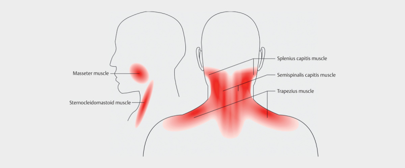
Purpose: to establish reference values for ultrasound shear-wave elastography for pericranial muscles in healthy individuals (m. trapezius, m. splenius capitis, m. semispinalis capitis, m. sternocleidomastoideus and m. masseter). Also to evaluate day-to-day variations in the shear-wave speeds and evaluate the effect of the pennation of the muscle fibers, ie scanning parallel or perpendicularly to the fibers.
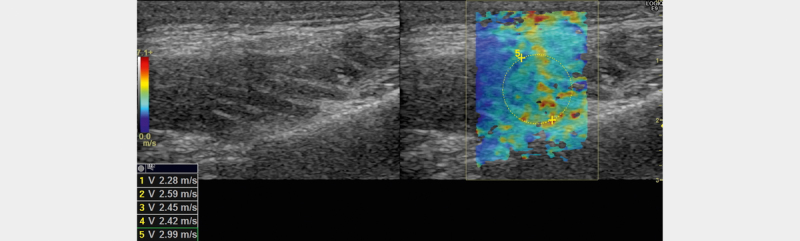
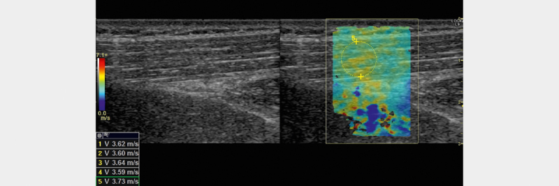
Materials and methods: 10 healthy individuals (5 males and 5 females) had their pericranial muscles examined with shear-wave elastography in two orthogonal planes on two different days for their dominant and non-dominant side. Mean shear wave speeds from 5 ROI's in each muscle, for each scan plane for the dominant and non-dominant side for the two days were calculated. The effect of the different parameters - muscle pennation, gender, dominant vs non-dominant side and day was evaluated.
Results: The effect of scan plane in relation to muscle pennation was statistically significant (p<0.0001). The mean shear-wave speed when scanning parallel to the muscle fibers was significantly higher than the mean shear-wave speed when scanning perpendicularly to the fibers. The day-to-day variation was statistically significant (p=0.0258), but not clinically relevant. Shear-wave speeds differed significantly between muscles. Mean shear wave speeds (m/s) for the muscles in the parallel plane were: for masseter 2.45 (SD:+/-0.25), semispinal 3.36 (SD:+/-0.75), splenius 3.04 (SD:+/-0.65), sternocleidomastoid 2.75 (SD:+/-0.23), trapezius 3.20 (SD:+/-0.27) and trapezius lateral 3.87 (SD:+/-3.87).
Conclusion: The shear wave speed variation depended on the direction of scanning. Shear wave elastography may be a method to evaluate muscle stiffness in patients suffering from chronic neck pain.


 分享
分享
 求助内容:
求助内容: 应助结果提醒方式:
 扫码关注我们
