Damiana Vulturar, Alexandru Fărcăşanu, Flaviu Turcu, Dan Boitor, Carmen Crivii
{"title":"小脑容量妊娠第二学期小脑的体积","authors":"Damiana Vulturar, Alexandru Fărcăşanu, Flaviu Turcu, Dan Boitor, Carmen Crivii","doi":"10.15386/cjmed-922","DOIUrl":null,"url":null,"abstract":"<p><strong>Background and aims: </strong>The cerebellum (\"little brain\"), the largest part of hind brain, lies in the posterior cranial fossa, beneath the occipital lobe and dorsal to the brainstem. It develops over a long period: it is one of the first structures in the brain to begin to differentiate, but one of the last to mature. The use of ultrasonography has significantly improved the evaluation of fetal growth and development and has permitted prenatal diagnosis of a variety of congenital malformations.The aim of our study was to evaluate the cerebellar growth and development using 2 different measuring techniques: microMRI and ultrasound technique. The cerebellum measurements were related to gestational age.</p><p><strong>Methods: </strong>We used 14 human fetuses corresponding to 15-28 gestational weeks, immersed in a 9% formalin solution. Magnetic Resonance Imaging (MRI) was performed by employing a Bruker BioSpec 70/16USR scanner (Bruker BioSpin MRI GmbH, Ettlingen, Germany), operated at 7.04 Tesla for cerebellar volume measurement. Ultrasonographic measurements of the cerebellum diameter were performed on 14 pregnant women, 15 - 28 gestational weeks. Ultrasound scan used 5-10 MHZ for transvaginal approach. Taking into consideration the values of the cerebellum dimensions and considering the general shape of the cerebellum as a transverse ellipsoid, the volume of the cerebellum was calculated by a mathematical formula for ellipsoid volume.</p><p><strong>Results: </strong>The study correlates the measurements from the microMRI study with the ultrasounds data and the results are superposable. Both established the exponential volume growth after the 22-23 GW. We used the ellipsoid volume formula for the cerebellar volume using the half of the three diameters of the cerebellum determined by ultrasound measurements:Cerebellar Volume = Ellipsoid volume = 3/4 π r1 r2 r3.</p><p><strong>Conclusion: </strong>There is a linear correlation between the microMRI measurements and ultrasound determinations. Based on all collected data we could apply an easy formula to calculate the volume of cerebellum, a useful criterion in the evaluation of the cerebellar development and the appreciation of the gestational age.</p>","PeriodicalId":91233,"journal":{"name":"Clujul medical (1957)","volume":"91 2","pages":"176-180"},"PeriodicalIF":0.0000,"publicationDate":"2018-01-01","publicationTypes":"Journal Article","fieldsOfStudy":null,"isOpenAccess":false,"openAccessPdf":"https://ftp.ncbi.nlm.nih.gov/pub/pmc/oa_pdf/18/53/cm-91-176.PMC5958982.pdf","citationCount":"7","resultStr":"{\"title\":\"The volume of the cerebellum in the second semester of gestation.\",\"authors\":\"Damiana Vulturar, Alexandru Fărcăşanu, Flaviu Turcu, Dan Boitor, Carmen Crivii\",\"doi\":\"10.15386/cjmed-922\",\"DOIUrl\":null,\"url\":null,\"abstract\":\"<p><strong>Background and aims: </strong>The cerebellum (\\\"little brain\\\"), the largest part of hind brain, lies in the posterior cranial fossa, beneath the occipital lobe and dorsal to the brainstem. It develops over a long period: it is one of the first structures in the brain to begin to differentiate, but one of the last to mature. The use of ultrasonography has significantly improved the evaluation of fetal growth and development and has permitted prenatal diagnosis of a variety of congenital malformations.The aim of our study was to evaluate the cerebellar growth and development using 2 different measuring techniques: microMRI and ultrasound technique. The cerebellum measurements were related to gestational age.</p><p><strong>Methods: </strong>We used 14 human fetuses corresponding to 15-28 gestational weeks, immersed in a 9% formalin solution. Magnetic Resonance Imaging (MRI) was performed by employing a Bruker BioSpec 70/16USR scanner (Bruker BioSpin MRI GmbH, Ettlingen, Germany), operated at 7.04 Tesla for cerebellar volume measurement. Ultrasonographic measurements of the cerebellum diameter were performed on 14 pregnant women, 15 - 28 gestational weeks. Ultrasound scan used 5-10 MHZ for transvaginal approach. Taking into consideration the values of the cerebellum dimensions and considering the general shape of the cerebellum as a transverse ellipsoid, the volume of the cerebellum was calculated by a mathematical formula for ellipsoid volume.</p><p><strong>Results: </strong>The study correlates the measurements from the microMRI study with the ultrasounds data and the results are superposable. Both established the exponential volume growth after the 22-23 GW. We used the ellipsoid volume formula for the cerebellar volume using the half of the three diameters of the cerebellum determined by ultrasound measurements:Cerebellar Volume = Ellipsoid volume = 3/4 π r1 r2 r3.</p><p><strong>Conclusion: </strong>There is a linear correlation between the microMRI measurements and ultrasound determinations. Based on all collected data we could apply an easy formula to calculate the volume of cerebellum, a useful criterion in the evaluation of the cerebellar development and the appreciation of the gestational age.</p>\",\"PeriodicalId\":91233,\"journal\":{\"name\":\"Clujul medical (1957)\",\"volume\":\"91 2\",\"pages\":\"176-180\"},\"PeriodicalIF\":0.0000,\"publicationDate\":\"2018-01-01\",\"publicationTypes\":\"Journal Article\",\"fieldsOfStudy\":null,\"isOpenAccess\":false,\"openAccessPdf\":\"https://ftp.ncbi.nlm.nih.gov/pub/pmc/oa_pdf/18/53/cm-91-176.PMC5958982.pdf\",\"citationCount\":\"7\",\"resultStr\":null,\"platform\":\"Semanticscholar\",\"paperid\":null,\"PeriodicalName\":\"Clujul medical (1957)\",\"FirstCategoryId\":\"1085\",\"ListUrlMain\":\"https://doi.org/10.15386/cjmed-922\",\"RegionNum\":0,\"RegionCategory\":null,\"ArticlePicture\":[],\"TitleCN\":null,\"AbstractTextCN\":null,\"PMCID\":null,\"EPubDate\":\"2018/4/25 0:00:00\",\"PubModel\":\"Epub\",\"JCR\":\"\",\"JCRName\":\"\",\"Score\":null,\"Total\":0}","platform":"Semanticscholar","paperid":null,"PeriodicalName":"Clujul medical (1957)","FirstCategoryId":"1085","ListUrlMain":"https://doi.org/10.15386/cjmed-922","RegionNum":0,"RegionCategory":null,"ArticlePicture":[],"TitleCN":null,"AbstractTextCN":null,"PMCID":null,"EPubDate":"2018/4/25 0:00:00","PubModel":"Epub","JCR":"","JCRName":"","Score":null,"Total":0}
The volume of the cerebellum in the second semester of gestation.
Background and aims: The cerebellum ("little brain"), the largest part of hind brain, lies in the posterior cranial fossa, beneath the occipital lobe and dorsal to the brainstem. It develops over a long period: it is one of the first structures in the brain to begin to differentiate, but one of the last to mature. The use of ultrasonography has significantly improved the evaluation of fetal growth and development and has permitted prenatal diagnosis of a variety of congenital malformations.The aim of our study was to evaluate the cerebellar growth and development using 2 different measuring techniques: microMRI and ultrasound technique. The cerebellum measurements were related to gestational age.
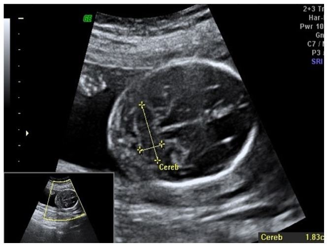
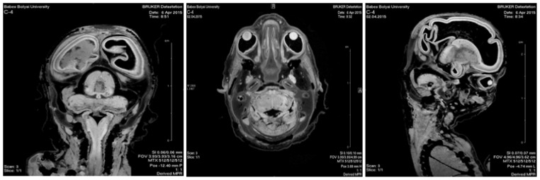
Methods: We used 14 human fetuses corresponding to 15-28 gestational weeks, immersed in a 9% formalin solution. Magnetic Resonance Imaging (MRI) was performed by employing a Bruker BioSpec 70/16USR scanner (Bruker BioSpin MRI GmbH, Ettlingen, Germany), operated at 7.04 Tesla for cerebellar volume measurement. Ultrasonographic measurements of the cerebellum diameter were performed on 14 pregnant women, 15 - 28 gestational weeks. Ultrasound scan used 5-10 MHZ for transvaginal approach. Taking into consideration the values of the cerebellum dimensions and considering the general shape of the cerebellum as a transverse ellipsoid, the volume of the cerebellum was calculated by a mathematical formula for ellipsoid volume.
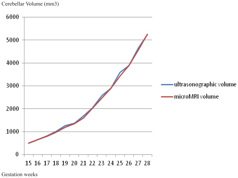
Results: The study correlates the measurements from the microMRI study with the ultrasounds data and the results are superposable. Both established the exponential volume growth after the 22-23 GW. We used the ellipsoid volume formula for the cerebellar volume using the half of the three diameters of the cerebellum determined by ultrasound measurements:Cerebellar Volume = Ellipsoid volume = 3/4 π r1 r2 r3.
Conclusion: There is a linear correlation between the microMRI measurements and ultrasound determinations. Based on all collected data we could apply an easy formula to calculate the volume of cerebellum, a useful criterion in the evaluation of the cerebellar development and the appreciation of the gestational age.


 分享
分享

 求助内容:
求助内容: 应助结果提醒方式:
 扫码关注我们
