Luxi Chen, Rosemary E Barnett, Martin Horstmann, Verena Bamberger, Lea Heberle, Nina Krebs, John K Colbourne, Rocío Gómez, Linda C Weiss
{"title":"水蚤滞育发育过程中有丝分裂活动模式和细胞骨架的变化。","authors":"Luxi Chen, Rosemary E Barnett, Martin Horstmann, Verena Bamberger, Lea Heberle, Nina Krebs, John K Colbourne, Rocío Gómez, Linda C Weiss","doi":"10.1186/s12860-018-0181-0","DOIUrl":null,"url":null,"abstract":"<p><strong>Background: </strong>Diapause is a form of dormancy that is genetically predetermined to allow animals to overcome harsh environmental conditions. It is induced by predictive environmental cues bringing cellular activity levels into a state of suspended animation. Entering diapause requires organismal, molecular and cellular adaptation to severely reduced energy flows. Cells must therefore have evolved strategies that prepare them for periods with limited metabolic resources. However, changes that occur on the (sub-)cellular level have not been thoroughly described.</p><p><strong>Results: </strong>We investigated mitotic activity and we monitored cytoskeletal network changes in successive stages of diapausing and non-diapausing Daphnia magna embryos using (immuno-)fluorescent labeling. We find that embryos destined to diapause show a delayed and 2.5x slower mitotic activity in comparison to continuously developing embryos. Development is halted when D. magna embryos reach ~ 3500 cells, whereupon mitotic activity is absent and cytoskeletal components are severely reduced, rendering diapause cells compact and condensed.</p><p><strong>Conclusion: </strong>In the initiation phase of diapause, the slower cell division rate points to prolonged interphase duration, preparing the cells for diapause maintenance. During diapause, cytoskeletal depletion and cellular condensation may be a means to save energy resources. Our data provide insights into the sub-cellular change of diapause in Daphnia.</p>","PeriodicalId":9051,"journal":{"name":"BMC Cell Biology","volume":" ","pages":"30"},"PeriodicalIF":0.0000,"publicationDate":"2018-12-29","publicationTypes":"Journal Article","fieldsOfStudy":null,"isOpenAccess":false,"openAccessPdf":"https://sci-hub-pdf.com/10.1186/s12860-018-0181-0","citationCount":"16","resultStr":"{\"title\":\"Mitotic activity patterns and cytoskeletal changes throughout the progression of diapause developmental program in Daphnia.\",\"authors\":\"Luxi Chen, Rosemary E Barnett, Martin Horstmann, Verena Bamberger, Lea Heberle, Nina Krebs, John K Colbourne, Rocío Gómez, Linda C Weiss\",\"doi\":\"10.1186/s12860-018-0181-0\",\"DOIUrl\":null,\"url\":null,\"abstract\":\"<p><strong>Background: </strong>Diapause is a form of dormancy that is genetically predetermined to allow animals to overcome harsh environmental conditions. It is induced by predictive environmental cues bringing cellular activity levels into a state of suspended animation. Entering diapause requires organismal, molecular and cellular adaptation to severely reduced energy flows. Cells must therefore have evolved strategies that prepare them for periods with limited metabolic resources. However, changes that occur on the (sub-)cellular level have not been thoroughly described.</p><p><strong>Results: </strong>We investigated mitotic activity and we monitored cytoskeletal network changes in successive stages of diapausing and non-diapausing Daphnia magna embryos using (immuno-)fluorescent labeling. We find that embryos destined to diapause show a delayed and 2.5x slower mitotic activity in comparison to continuously developing embryos. Development is halted when D. magna embryos reach ~ 3500 cells, whereupon mitotic activity is absent and cytoskeletal components are severely reduced, rendering diapause cells compact and condensed.</p><p><strong>Conclusion: </strong>In the initiation phase of diapause, the slower cell division rate points to prolonged interphase duration, preparing the cells for diapause maintenance. During diapause, cytoskeletal depletion and cellular condensation may be a means to save energy resources. Our data provide insights into the sub-cellular change of diapause in Daphnia.</p>\",\"PeriodicalId\":9051,\"journal\":{\"name\":\"BMC Cell Biology\",\"volume\":\" \",\"pages\":\"30\"},\"PeriodicalIF\":0.0000,\"publicationDate\":\"2018-12-29\",\"publicationTypes\":\"Journal Article\",\"fieldsOfStudy\":null,\"isOpenAccess\":false,\"openAccessPdf\":\"https://sci-hub-pdf.com/10.1186/s12860-018-0181-0\",\"citationCount\":\"16\",\"resultStr\":null,\"platform\":\"Semanticscholar\",\"paperid\":null,\"PeriodicalName\":\"BMC Cell Biology\",\"FirstCategoryId\":\"1085\",\"ListUrlMain\":\"https://doi.org/10.1186/s12860-018-0181-0\",\"RegionNum\":0,\"RegionCategory\":null,\"ArticlePicture\":[],\"TitleCN\":null,\"AbstractTextCN\":null,\"PMCID\":null,\"EPubDate\":\"\",\"PubModel\":\"\",\"JCR\":\"Q1\",\"JCRName\":\"Biochemistry, Genetics and Molecular Biology\",\"Score\":null,\"Total\":0}","platform":"Semanticscholar","paperid":null,"PeriodicalName":"BMC Cell Biology","FirstCategoryId":"1085","ListUrlMain":"https://doi.org/10.1186/s12860-018-0181-0","RegionNum":0,"RegionCategory":null,"ArticlePicture":[],"TitleCN":null,"AbstractTextCN":null,"PMCID":null,"EPubDate":"","PubModel":"","JCR":"Q1","JCRName":"Biochemistry, Genetics and Molecular Biology","Score":null,"Total":0}
Mitotic activity patterns and cytoskeletal changes throughout the progression of diapause developmental program in Daphnia.
Background: Diapause is a form of dormancy that is genetically predetermined to allow animals to overcome harsh environmental conditions. It is induced by predictive environmental cues bringing cellular activity levels into a state of suspended animation. Entering diapause requires organismal, molecular and cellular adaptation to severely reduced energy flows. Cells must therefore have evolved strategies that prepare them for periods with limited metabolic resources. However, changes that occur on the (sub-)cellular level have not been thoroughly described.
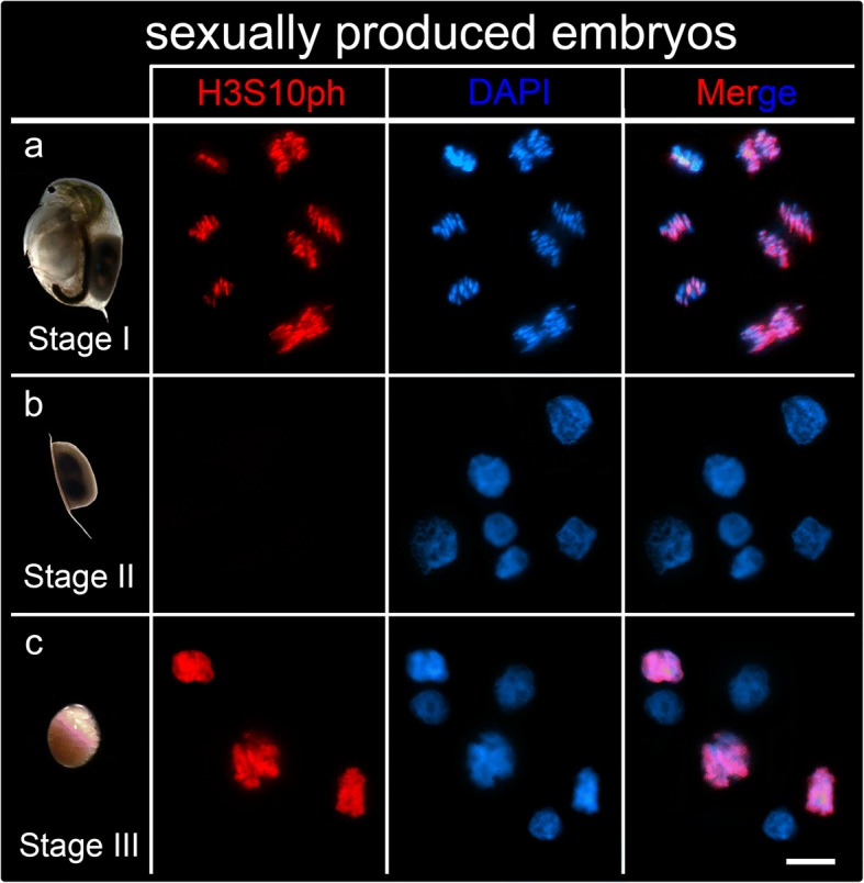
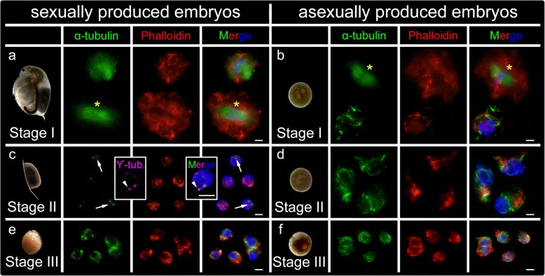
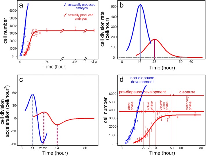
Results: We investigated mitotic activity and we monitored cytoskeletal network changes in successive stages of diapausing and non-diapausing Daphnia magna embryos using (immuno-)fluorescent labeling. We find that embryos destined to diapause show a delayed and 2.5x slower mitotic activity in comparison to continuously developing embryos. Development is halted when D. magna embryos reach ~ 3500 cells, whereupon mitotic activity is absent and cytoskeletal components are severely reduced, rendering diapause cells compact and condensed.
Conclusion: In the initiation phase of diapause, the slower cell division rate points to prolonged interphase duration, preparing the cells for diapause maintenance. During diapause, cytoskeletal depletion and cellular condensation may be a means to save energy resources. Our data provide insights into the sub-cellular change of diapause in Daphnia.
期刊介绍:
BMC Molecular and Cell Biology, formerly known as BMC Cell Biology, is an open access journal that considers articles on all aspects of both eukaryotic and prokaryotic cell and molecular biology, including structural and functional cell biology, DNA and RNA in a cellular context and biochemistry, as well as research using both the experimental and theoretical aspects of physics to study biological processes and investigations into the structure of biological macromolecules.


 分享
分享
 求助内容:
求助内容: 应助结果提醒方式:
 扫码关注我们
