Francisco Barbosa De Araujo Neto, Camila Corona De Godoy Bueno, Liege Tambelini Gomes, Daniela Alejandra Ortiz Navas, Mark Wanderley, Stefanie Gallotti Borges Carneiro, Rita Karine Veras Gomes De Mello, Laura Mendes Coura, Larissa Sayuri Missumi, Henrique Durante, Ricardo Francisco Cintra Zagatti, Márcio Valente Yamada Sawamura
{"title":"罕见的隐球菌感染的诊断挑战。","authors":"Francisco Barbosa De Araujo Neto, Camila Corona De Godoy Bueno, Liege Tambelini Gomes, Daniela Alejandra Ortiz Navas, Mark Wanderley, Stefanie Gallotti Borges Carneiro, Rita Karine Veras Gomes De Mello, Laura Mendes Coura, Larissa Sayuri Missumi, Henrique Durante, Ricardo Francisco Cintra Zagatti, Márcio Valente Yamada Sawamura","doi":"10.1155/2019/5970648","DOIUrl":null,"url":null,"abstract":"<p><p>Cryptococcal infection results from inhalation of fungal spores and usually is confined to the lungs, but may disseminate systemically. Radiologically, cryptococcal infection has multiple forms of presentation. The diagnosis is usually based on fungal isolation from cultured clinical specimens. Long term antifungal therapy is recommended, but surgical procedures may eventually be necessary when large thoracic symptomatic masses are present. We report a case of a 41-year-old male, immunocompetent, investigating a palpable mass in the left supraclavicular region associated with unintentional weight loss over the last three months. He also reported chest pain in this period. Chest X-ray, ultrasonography, and computed tomography were performed, which diagnosed a mediastinal and left supraclavicular mass, interpreted as lymph node conglomerates of unknown etiology. He also underwent a biopsy of the left supraclavicular mass for etiological determination by histopathology, which confirmed cryptococcosis infection. Although very infrequent, mediastinal cryptococcal infection (simulating masses) is a challenging but important differential diagnosis of benign and malignant lesions, since its treatment is usually clinical.</p>","PeriodicalId":30326,"journal":{"name":"Case Reports in Radiology","volume":" ","pages":"5970648"},"PeriodicalIF":0.0000,"publicationDate":"2019-01-02","publicationTypes":"Journal Article","fieldsOfStudy":null,"isOpenAccess":false,"openAccessPdf":"https://sci-hub-pdf.com/10.1155/2019/5970648","citationCount":"2","resultStr":"{\"title\":\"The Diagnostic Challenge of an Infrequent Spectrum of <i>Cryptococcus</i> Infection.\",\"authors\":\"Francisco Barbosa De Araujo Neto, Camila Corona De Godoy Bueno, Liege Tambelini Gomes, Daniela Alejandra Ortiz Navas, Mark Wanderley, Stefanie Gallotti Borges Carneiro, Rita Karine Veras Gomes De Mello, Laura Mendes Coura, Larissa Sayuri Missumi, Henrique Durante, Ricardo Francisco Cintra Zagatti, Márcio Valente Yamada Sawamura\",\"doi\":\"10.1155/2019/5970648\",\"DOIUrl\":null,\"url\":null,\"abstract\":\"<p><p>Cryptococcal infection results from inhalation of fungal spores and usually is confined to the lungs, but may disseminate systemically. Radiologically, cryptococcal infection has multiple forms of presentation. The diagnosis is usually based on fungal isolation from cultured clinical specimens. Long term antifungal therapy is recommended, but surgical procedures may eventually be necessary when large thoracic symptomatic masses are present. We report a case of a 41-year-old male, immunocompetent, investigating a palpable mass in the left supraclavicular region associated with unintentional weight loss over the last three months. He also reported chest pain in this period. Chest X-ray, ultrasonography, and computed tomography were performed, which diagnosed a mediastinal and left supraclavicular mass, interpreted as lymph node conglomerates of unknown etiology. He also underwent a biopsy of the left supraclavicular mass for etiological determination by histopathology, which confirmed cryptococcosis infection. Although very infrequent, mediastinal cryptococcal infection (simulating masses) is a challenging but important differential diagnosis of benign and malignant lesions, since its treatment is usually clinical.</p>\",\"PeriodicalId\":30326,\"journal\":{\"name\":\"Case Reports in Radiology\",\"volume\":\" \",\"pages\":\"5970648\"},\"PeriodicalIF\":0.0000,\"publicationDate\":\"2019-01-02\",\"publicationTypes\":\"Journal Article\",\"fieldsOfStudy\":null,\"isOpenAccess\":false,\"openAccessPdf\":\"https://sci-hub-pdf.com/10.1155/2019/5970648\",\"citationCount\":\"2\",\"resultStr\":null,\"platform\":\"Semanticscholar\",\"paperid\":null,\"PeriodicalName\":\"Case Reports in Radiology\",\"FirstCategoryId\":\"1085\",\"ListUrlMain\":\"https://doi.org/10.1155/2019/5970648\",\"RegionNum\":0,\"RegionCategory\":null,\"ArticlePicture\":[],\"TitleCN\":null,\"AbstractTextCN\":null,\"PMCID\":null,\"EPubDate\":\"2019/1/1 0:00:00\",\"PubModel\":\"eCollection\",\"JCR\":\"\",\"JCRName\":\"\",\"Score\":null,\"Total\":0}","platform":"Semanticscholar","paperid":null,"PeriodicalName":"Case Reports in Radiology","FirstCategoryId":"1085","ListUrlMain":"https://doi.org/10.1155/2019/5970648","RegionNum":0,"RegionCategory":null,"ArticlePicture":[],"TitleCN":null,"AbstractTextCN":null,"PMCID":null,"EPubDate":"2019/1/1 0:00:00","PubModel":"eCollection","JCR":"","JCRName":"","Score":null,"Total":0}
The Diagnostic Challenge of an Infrequent Spectrum of Cryptococcus Infection.
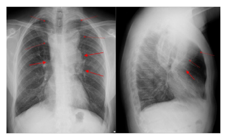
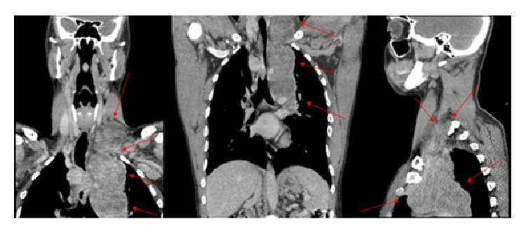
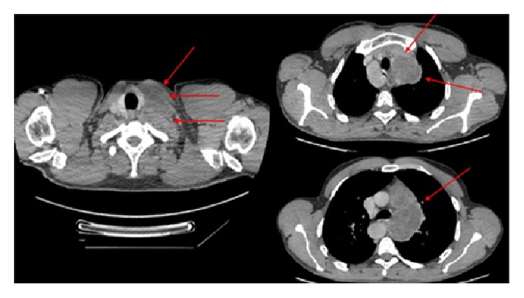
Cryptococcal infection results from inhalation of fungal spores and usually is confined to the lungs, but may disseminate systemically. Radiologically, cryptococcal infection has multiple forms of presentation. The diagnosis is usually based on fungal isolation from cultured clinical specimens. Long term antifungal therapy is recommended, but surgical procedures may eventually be necessary when large thoracic symptomatic masses are present. We report a case of a 41-year-old male, immunocompetent, investigating a palpable mass in the left supraclavicular region associated with unintentional weight loss over the last three months. He also reported chest pain in this period. Chest X-ray, ultrasonography, and computed tomography were performed, which diagnosed a mediastinal and left supraclavicular mass, interpreted as lymph node conglomerates of unknown etiology. He also underwent a biopsy of the left supraclavicular mass for etiological determination by histopathology, which confirmed cryptococcosis infection. Although very infrequent, mediastinal cryptococcal infection (simulating masses) is a challenging but important differential diagnosis of benign and malignant lesions, since its treatment is usually clinical.


 分享
分享

 求助内容:
求助内容: 应助结果提醒方式:
 扫码关注我们
