Houman Mirzaalian Dastjerdi, Dominique Töpfer, Stefan J Rupitsch, Andreas Maier
{"title":"用二维和三维算法测量皮肤病变表面积。","authors":"Houman Mirzaalian Dastjerdi, Dominique Töpfer, Stefan J Rupitsch, Andreas Maier","doi":"10.1155/2019/4035148","DOIUrl":null,"url":null,"abstract":"<p><strong>Purpose: </strong>The treatment of skin lesions of various kinds is a common task in clinical routine. Apart from wound care, the assessment of treatment efficacy plays an important role. In this paper, we present a new approach to measure the skin lesion surface in two and three dimensions.</p><p><strong>Methods: </strong>For the 2D approach, a single photo containing a flexible paper ruler is taken. After semi-automatic segmentation of the lesion, evaluation is based on local scale estimation using the ruler. For the 3D approach, reconstruction is based on Structure from Motion. Roughly outlining the region of interest around the lesion is required for both methods.</p><p><strong>Results: </strong>The measurement evaluation was performed on 117 phantom images and five phantom videos for 2D and 3D approach, respectively. We found an absolute error of 0.99±1.18 cm<sup>2</sup> and a relative error 9.89± 9.31% for 2D. These errors are <1 cm<sup>2</sup> and <5% for five test phantoms in our 3D case. As expected, the error of 2D surface area measurement increased by approximately 10% for wounds on the bent surface compared to wounds on the flat surface. Using our method, the only user interaction is to roughly outline the region of interest around the lesion.</p><p><strong>Conclusions: </strong>We developed a new wound segmentation and surface area measurement technique for skin lesions even on a bent surface. The 2D technique provides the user with a fast, user-friendly segmentation and measurement tool with reasonable accuracy for home care assessment of treatment. For 3D only preliminary results could be provided. Measurements were only based on phantoms and have to be repeated with real clinical data.</p>","PeriodicalId":47063,"journal":{"name":"International Journal of Biomedical Imaging","volume":" ","pages":"4035148"},"PeriodicalIF":1.3000,"publicationDate":"2019-01-15","publicationTypes":"Journal Article","fieldsOfStudy":null,"isOpenAccess":false,"openAccessPdf":"https://sci-hub-pdf.com/10.1155/2019/4035148","citationCount":"17","resultStr":"{\"title\":\"Measuring Surface Area of Skin Lesions with 2D and 3D Algorithms.\",\"authors\":\"Houman Mirzaalian Dastjerdi, Dominique Töpfer, Stefan J Rupitsch, Andreas Maier\",\"doi\":\"10.1155/2019/4035148\",\"DOIUrl\":null,\"url\":null,\"abstract\":\"<p><strong>Purpose: </strong>The treatment of skin lesions of various kinds is a common task in clinical routine. Apart from wound care, the assessment of treatment efficacy plays an important role. In this paper, we present a new approach to measure the skin lesion surface in two and three dimensions.</p><p><strong>Methods: </strong>For the 2D approach, a single photo containing a flexible paper ruler is taken. After semi-automatic segmentation of the lesion, evaluation is based on local scale estimation using the ruler. For the 3D approach, reconstruction is based on Structure from Motion. Roughly outlining the region of interest around the lesion is required for both methods.</p><p><strong>Results: </strong>The measurement evaluation was performed on 117 phantom images and five phantom videos for 2D and 3D approach, respectively. We found an absolute error of 0.99±1.18 cm<sup>2</sup> and a relative error 9.89± 9.31% for 2D. These errors are <1 cm<sup>2</sup> and <5% for five test phantoms in our 3D case. As expected, the error of 2D surface area measurement increased by approximately 10% for wounds on the bent surface compared to wounds on the flat surface. Using our method, the only user interaction is to roughly outline the region of interest around the lesion.</p><p><strong>Conclusions: </strong>We developed a new wound segmentation and surface area measurement technique for skin lesions even on a bent surface. The 2D technique provides the user with a fast, user-friendly segmentation and measurement tool with reasonable accuracy for home care assessment of treatment. For 3D only preliminary results could be provided. Measurements were only based on phantoms and have to be repeated with real clinical data.</p>\",\"PeriodicalId\":47063,\"journal\":{\"name\":\"International Journal of Biomedical Imaging\",\"volume\":\" \",\"pages\":\"4035148\"},\"PeriodicalIF\":1.3000,\"publicationDate\":\"2019-01-15\",\"publicationTypes\":\"Journal Article\",\"fieldsOfStudy\":null,\"isOpenAccess\":false,\"openAccessPdf\":\"https://sci-hub-pdf.com/10.1155/2019/4035148\",\"citationCount\":\"17\",\"resultStr\":null,\"platform\":\"Semanticscholar\",\"paperid\":null,\"PeriodicalName\":\"International Journal of Biomedical Imaging\",\"FirstCategoryId\":\"1085\",\"ListUrlMain\":\"https://doi.org/10.1155/2019/4035148\",\"RegionNum\":0,\"RegionCategory\":null,\"ArticlePicture\":[],\"TitleCN\":null,\"AbstractTextCN\":null,\"PMCID\":null,\"EPubDate\":\"2019/1/1 0:00:00\",\"PubModel\":\"eCollection\",\"JCR\":\"Q2\",\"JCRName\":\"ENGINEERING, BIOMEDICAL\",\"Score\":null,\"Total\":0}","platform":"Semanticscholar","paperid":null,"PeriodicalName":"International Journal of Biomedical Imaging","FirstCategoryId":"1085","ListUrlMain":"https://doi.org/10.1155/2019/4035148","RegionNum":0,"RegionCategory":null,"ArticlePicture":[],"TitleCN":null,"AbstractTextCN":null,"PMCID":null,"EPubDate":"2019/1/1 0:00:00","PubModel":"eCollection","JCR":"Q2","JCRName":"ENGINEERING, BIOMEDICAL","Score":null,"Total":0}
Measuring Surface Area of Skin Lesions with 2D and 3D Algorithms.
Purpose: The treatment of skin lesions of various kinds is a common task in clinical routine. Apart from wound care, the assessment of treatment efficacy plays an important role. In this paper, we present a new approach to measure the skin lesion surface in two and three dimensions.
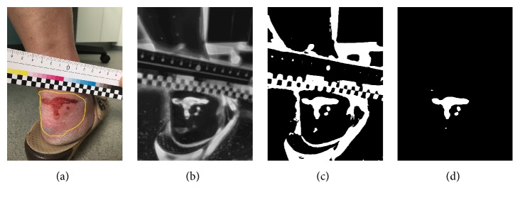
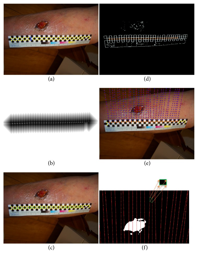
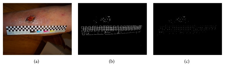
Methods: For the 2D approach, a single photo containing a flexible paper ruler is taken. After semi-automatic segmentation of the lesion, evaluation is based on local scale estimation using the ruler. For the 3D approach, reconstruction is based on Structure from Motion. Roughly outlining the region of interest around the lesion is required for both methods.
Results: The measurement evaluation was performed on 117 phantom images and five phantom videos for 2D and 3D approach, respectively. We found an absolute error of 0.99±1.18 cm2 and a relative error 9.89± 9.31% for 2D. These errors are <1 cm2 and <5% for five test phantoms in our 3D case. As expected, the error of 2D surface area measurement increased by approximately 10% for wounds on the bent surface compared to wounds on the flat surface. Using our method, the only user interaction is to roughly outline the region of interest around the lesion.
Conclusions: We developed a new wound segmentation and surface area measurement technique for skin lesions even on a bent surface. The 2D technique provides the user with a fast, user-friendly segmentation and measurement tool with reasonable accuracy for home care assessment of treatment. For 3D only preliminary results could be provided. Measurements were only based on phantoms and have to be repeated with real clinical data.
期刊介绍:
The International Journal of Biomedical Imaging is managed by a board of editors comprising internationally renowned active researchers. The journal is freely accessible online and also offered for purchase in print format. It employs a web-based review system to ensure swift turnaround times while maintaining high standards. In addition to regular issues, special issues are organized by guest editors. The subject areas covered include (but are not limited to):
Digital radiography and tomosynthesis
X-ray computed tomography (CT)
Magnetic resonance imaging (MRI)
Single photon emission computed tomography (SPECT)
Positron emission tomography (PET)
Ultrasound imaging
Diffuse optical tomography, coherence, fluorescence, bioluminescence tomography, impedance tomography
Neutron imaging for biomedical applications
Magnetic and optical spectroscopy, and optical biopsy
Optical, electron, scanning tunneling/atomic force microscopy
Small animal imaging
Functional, cellular, and molecular imaging
Imaging assays for screening and molecular analysis
Microarray image analysis and bioinformatics
Emerging biomedical imaging techniques
Imaging modality fusion
Biomedical imaging instrumentation
Biomedical image processing, pattern recognition, and analysis
Biomedical image visualization, compression, transmission, and storage
Imaging and modeling related to systems biology and systems biomedicine
Applied mathematics, applied physics, and chemistry related to biomedical imaging
Grid-enabling technology for biomedical imaging and informatics


 分享
分享
 求助内容:
求助内容: 应助结果提醒方式:
 扫码关注我们
