{"title":"应用扩散张量成像和纤维跟踪的组合立体技术评估臂丛。","authors":"Niyazi Acer, Mehmet Turgut","doi":"10.1055/s-0039-1687913","DOIUrl":null,"url":null,"abstract":"<p><p><b>Background</b> Brachial plexus (BP) is composed of intercommunications among the ventral roots of the nerves C5, C6, C7, C8, and T1 in the neck. The in vivo and in vitro evaluation of axons of the peripheral nervous system is performed using different techniques. Recently, many studies describing the application of fiber tractography and stereological axon number estimation to peripheral nerves have been published. <b>Methods</b> Various quantitative parameters of nerve fibers, including axon number, density, axonal area, and myelin thickness, can be estimated using stereological techniques. In vivo three-dimensional reconstruction of axons of BP can be visualized using a combined technique of diffusion tensor imaging (DTI) and fiber tracking with the potential to evaluate nerve fiber content. <b>Conclusion</b> It is concluded that terminal branches of BP can be successfully visualized using DTI, which is a highly reproducible method for the evaluation of BP as it shows anatomical and functional features of neural structures. We believe that quantitative morphological findings obtained from BP will be useful for new experimental, developmental, and pathological studies in the future.</p>","PeriodicalId":15280,"journal":{"name":"Journal of Brachial Plexus and Peripheral Nerve Injury","volume":"14 1","pages":"e16-e23"},"PeriodicalIF":1.0000,"publicationDate":"2019-06-12","publicationTypes":"Journal Article","fieldsOfStudy":null,"isOpenAccess":false,"openAccessPdf":"https://sci-hub-pdf.com/10.1055/s-0039-1687913","citationCount":"3","resultStr":"{\"title\":\"Evaluation of Brachial Plexus Using Combined Stereological Techniques of Diffusion Tensor Imaging and Fiber Tracking.\",\"authors\":\"Niyazi Acer, Mehmet Turgut\",\"doi\":\"10.1055/s-0039-1687913\",\"DOIUrl\":null,\"url\":null,\"abstract\":\"<p><p><b>Background</b> Brachial plexus (BP) is composed of intercommunications among the ventral roots of the nerves C5, C6, C7, C8, and T1 in the neck. The in vivo and in vitro evaluation of axons of the peripheral nervous system is performed using different techniques. Recently, many studies describing the application of fiber tractography and stereological axon number estimation to peripheral nerves have been published. <b>Methods</b> Various quantitative parameters of nerve fibers, including axon number, density, axonal area, and myelin thickness, can be estimated using stereological techniques. In vivo three-dimensional reconstruction of axons of BP can be visualized using a combined technique of diffusion tensor imaging (DTI) and fiber tracking with the potential to evaluate nerve fiber content. <b>Conclusion</b> It is concluded that terminal branches of BP can be successfully visualized using DTI, which is a highly reproducible method for the evaluation of BP as it shows anatomical and functional features of neural structures. We believe that quantitative morphological findings obtained from BP will be useful for new experimental, developmental, and pathological studies in the future.</p>\",\"PeriodicalId\":15280,\"journal\":{\"name\":\"Journal of Brachial Plexus and Peripheral Nerve Injury\",\"volume\":\"14 1\",\"pages\":\"e16-e23\"},\"PeriodicalIF\":1.0000,\"publicationDate\":\"2019-06-12\",\"publicationTypes\":\"Journal Article\",\"fieldsOfStudy\":null,\"isOpenAccess\":false,\"openAccessPdf\":\"https://sci-hub-pdf.com/10.1055/s-0039-1687913\",\"citationCount\":\"3\",\"resultStr\":null,\"platform\":\"Semanticscholar\",\"paperid\":null,\"PeriodicalName\":\"Journal of Brachial Plexus and Peripheral Nerve Injury\",\"FirstCategoryId\":\"1085\",\"ListUrlMain\":\"https://doi.org/10.1055/s-0039-1687913\",\"RegionNum\":0,\"RegionCategory\":null,\"ArticlePicture\":[],\"TitleCN\":null,\"AbstractTextCN\":null,\"PMCID\":null,\"EPubDate\":\"2019/1/1 0:00:00\",\"PubModel\":\"eCollection\",\"JCR\":\"Q4\",\"JCRName\":\"CLINICAL NEUROLOGY\",\"Score\":null,\"Total\":0}","platform":"Semanticscholar","paperid":null,"PeriodicalName":"Journal of Brachial Plexus and Peripheral Nerve Injury","FirstCategoryId":"1085","ListUrlMain":"https://doi.org/10.1055/s-0039-1687913","RegionNum":0,"RegionCategory":null,"ArticlePicture":[],"TitleCN":null,"AbstractTextCN":null,"PMCID":null,"EPubDate":"2019/1/1 0:00:00","PubModel":"eCollection","JCR":"Q4","JCRName":"CLINICAL NEUROLOGY","Score":null,"Total":0}
Evaluation of Brachial Plexus Using Combined Stereological Techniques of Diffusion Tensor Imaging and Fiber Tracking.
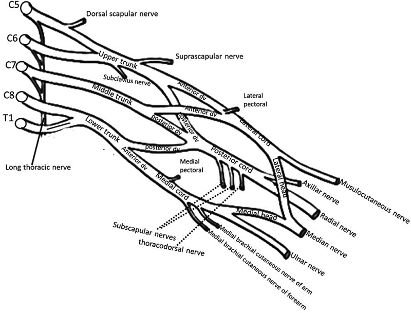
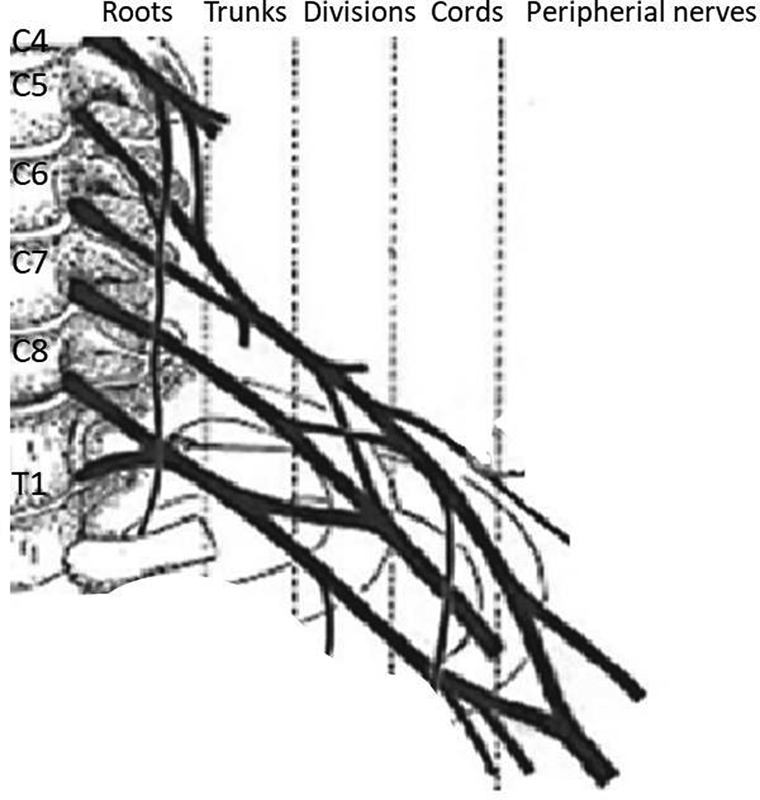
Background Brachial plexus (BP) is composed of intercommunications among the ventral roots of the nerves C5, C6, C7, C8, and T1 in the neck. The in vivo and in vitro evaluation of axons of the peripheral nervous system is performed using different techniques. Recently, many studies describing the application of fiber tractography and stereological axon number estimation to peripheral nerves have been published. Methods Various quantitative parameters of nerve fibers, including axon number, density, axonal area, and myelin thickness, can be estimated using stereological techniques. In vivo three-dimensional reconstruction of axons of BP can be visualized using a combined technique of diffusion tensor imaging (DTI) and fiber tracking with the potential to evaluate nerve fiber content. Conclusion It is concluded that terminal branches of BP can be successfully visualized using DTI, which is a highly reproducible method for the evaluation of BP as it shows anatomical and functional features of neural structures. We believe that quantitative morphological findings obtained from BP will be useful for new experimental, developmental, and pathological studies in the future.
期刊介绍:
JBPPNI is an open access, peer-reviewed online journal that will encompass all aspects of basic and clinical research findings, in the area of brachial plexus and peripheral nerve injury. Injury in this context refers to congenital, inflammatory, traumatic, degenerative and neoplastic processes, including neurofibromatosis. Papers on diagnostic and imaging aspects of the peripheral nervous system are welcomed as well. The peripheral nervous system is unique in its complexity and scope of influence. There are areas of interest in the anatomy, physiology, metabolism, phylogeny, and limb growth tropism of peripheral nerves.


 分享
分享
 求助内容:
求助内容: 应助结果提醒方式:
 扫码关注我们
