Michael R Gardner, Ayesha S Rahman, Thomas E Milner, Henry G Rylander
{"title":"缺氧小鼠视网膜模型的散射-角度分辨光学相干断层成像。","authors":"Michael R Gardner, Ayesha S Rahman, Thomas E Milner, Henry G Rylander","doi":"10.1177/1179069519837564","DOIUrl":null,"url":null,"abstract":"<p><p>Several studies have noted a correlation between retinal degeneration and traumatic encephalopathy (TE) making the retina a leading candidate for detection and assessment. Scattering-angle-resolved optical coherence tomography (SAR-OCT) is a candidate imaging modality to detect sub-resolution changes in retinal microstructure. SAR-OCT images of murine retinas that experience a hypoxic insult-euthanasia by isoflurane overdose-are presented. A total of 4 SAR-OCT measurement parameters are reported in 6 longitudinal experiments: blood flow volume fraction, total retinal thickness, reflectance index, and scattering angle. As each mouse expires, blood flow volume fraction decreases, total retinal thickness increases, reflectance index decreases, and scattering angle diversity increases. Contribution of the retinal vasculature to scattering angle diversity is discussed. Results of this study suggest the utility of SAR-OCT to measure TE using scattering angle diversity contrast in the retina.</p>","PeriodicalId":15817,"journal":{"name":"Journal of Experimental Neuroscience","volume":"13 ","pages":"1179069519837564"},"PeriodicalIF":0.0000,"publicationDate":"2019-03-27","publicationTypes":"Journal Article","fieldsOfStudy":null,"isOpenAccess":false,"openAccessPdf":"https://ftp.ncbi.nlm.nih.gov/pub/pmc/oa_pdf/ec/7c/10.1177_1179069519837564.PMC6440039.pdf","citationCount":"0","resultStr":"{\"title\":\"Scattering-Angle-Resolved Optical Coherence Tomography of a Hypoxic Mouse Retina Model.\",\"authors\":\"Michael R Gardner, Ayesha S Rahman, Thomas E Milner, Henry G Rylander\",\"doi\":\"10.1177/1179069519837564\",\"DOIUrl\":null,\"url\":null,\"abstract\":\"<p><p>Several studies have noted a correlation between retinal degeneration and traumatic encephalopathy (TE) making the retina a leading candidate for detection and assessment. Scattering-angle-resolved optical coherence tomography (SAR-OCT) is a candidate imaging modality to detect sub-resolution changes in retinal microstructure. SAR-OCT images of murine retinas that experience a hypoxic insult-euthanasia by isoflurane overdose-are presented. A total of 4 SAR-OCT measurement parameters are reported in 6 longitudinal experiments: blood flow volume fraction, total retinal thickness, reflectance index, and scattering angle. As each mouse expires, blood flow volume fraction decreases, total retinal thickness increases, reflectance index decreases, and scattering angle diversity increases. Contribution of the retinal vasculature to scattering angle diversity is discussed. Results of this study suggest the utility of SAR-OCT to measure TE using scattering angle diversity contrast in the retina.</p>\",\"PeriodicalId\":15817,\"journal\":{\"name\":\"Journal of Experimental Neuroscience\",\"volume\":\"13 \",\"pages\":\"1179069519837564\"},\"PeriodicalIF\":0.0000,\"publicationDate\":\"2019-03-27\",\"publicationTypes\":\"Journal Article\",\"fieldsOfStudy\":null,\"isOpenAccess\":false,\"openAccessPdf\":\"https://ftp.ncbi.nlm.nih.gov/pub/pmc/oa_pdf/ec/7c/10.1177_1179069519837564.PMC6440039.pdf\",\"citationCount\":\"0\",\"resultStr\":null,\"platform\":\"Semanticscholar\",\"paperid\":null,\"PeriodicalName\":\"Journal of Experimental Neuroscience\",\"FirstCategoryId\":\"1085\",\"ListUrlMain\":\"https://doi.org/10.1177/1179069519837564\",\"RegionNum\":0,\"RegionCategory\":null,\"ArticlePicture\":[],\"TitleCN\":null,\"AbstractTextCN\":null,\"PMCID\":null,\"EPubDate\":\"2019/1/1 0:00:00\",\"PubModel\":\"eCollection\",\"JCR\":\"\",\"JCRName\":\"\",\"Score\":null,\"Total\":0}","platform":"Semanticscholar","paperid":null,"PeriodicalName":"Journal of Experimental Neuroscience","FirstCategoryId":"1085","ListUrlMain":"https://doi.org/10.1177/1179069519837564","RegionNum":0,"RegionCategory":null,"ArticlePicture":[],"TitleCN":null,"AbstractTextCN":null,"PMCID":null,"EPubDate":"2019/1/1 0:00:00","PubModel":"eCollection","JCR":"","JCRName":"","Score":null,"Total":0}
Scattering-Angle-Resolved Optical Coherence Tomography of a Hypoxic Mouse Retina Model.
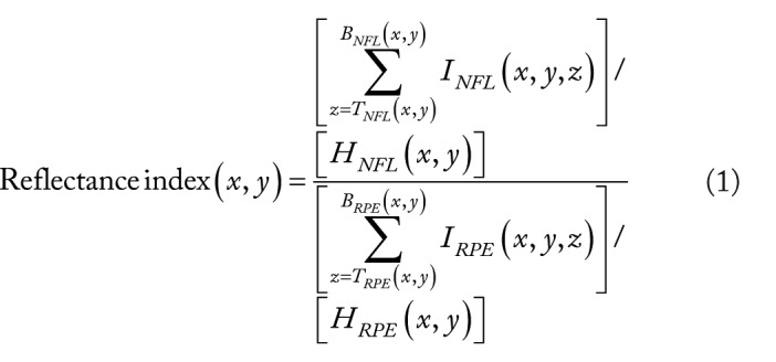
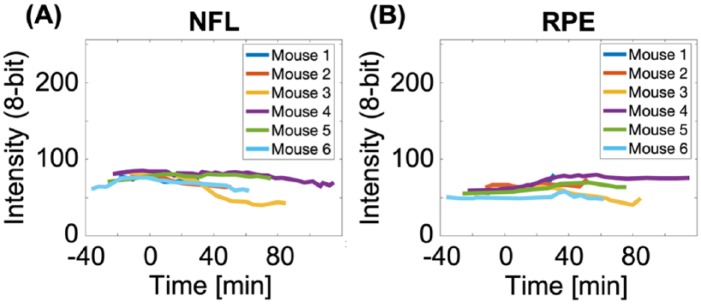
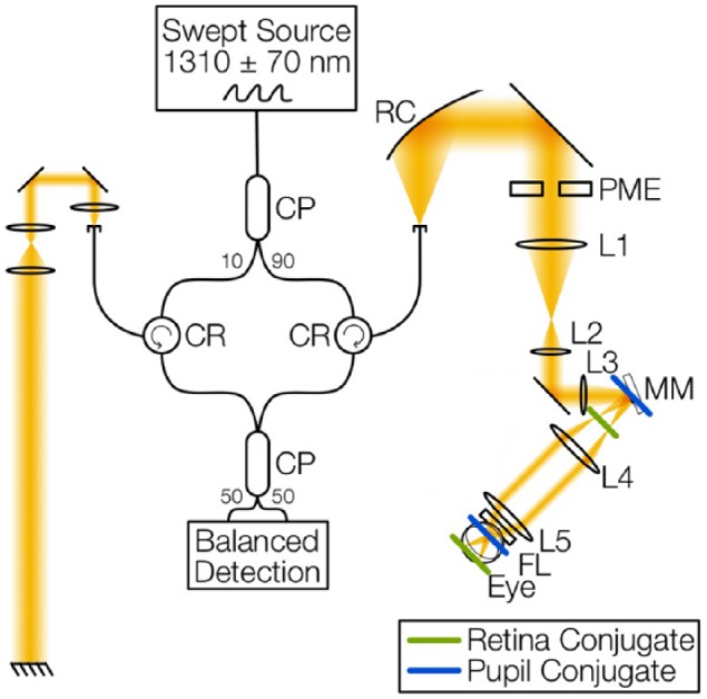
Several studies have noted a correlation between retinal degeneration and traumatic encephalopathy (TE) making the retina a leading candidate for detection and assessment. Scattering-angle-resolved optical coherence tomography (SAR-OCT) is a candidate imaging modality to detect sub-resolution changes in retinal microstructure. SAR-OCT images of murine retinas that experience a hypoxic insult-euthanasia by isoflurane overdose-are presented. A total of 4 SAR-OCT measurement parameters are reported in 6 longitudinal experiments: blood flow volume fraction, total retinal thickness, reflectance index, and scattering angle. As each mouse expires, blood flow volume fraction decreases, total retinal thickness increases, reflectance index decreases, and scattering angle diversity increases. Contribution of the retinal vasculature to scattering angle diversity is discussed. Results of this study suggest the utility of SAR-OCT to measure TE using scattering angle diversity contrast in the retina.


 分享
分享
 求助内容:
求助内容: 应助结果提醒方式:
 扫码关注我们
