Eleni-Anastasia Triantafyllou, Ilias Mylonis, George Simos, Efrosyni Paraskeva
{"title":"缺氧诱导肝癌细胞中不依赖炎症刺激和NF-κΒ通路的促纤维化和纤维化标记基因。","authors":"Eleni-Anastasia Triantafyllou, Ilias Mylonis, George Simos, Efrosyni Paraskeva","doi":"10.2147/HP.S235967","DOIUrl":null,"url":null,"abstract":"<p><p>Hypoxia and its key mediators hypoxia inducible Factors (HIFs) are implicated in the development of liver diseases of diverse etiologies, often in interplay with inflammatory mediators. We investigated the interplay between hypoxia and proinflammatory mediators in the development of liver fibrosis, using human hepatocellular carcinoma Huh7 cells as a model. Treatment of Huh7 with DMOG or under hypoxia, induced HIF-1α protein levels and the expression of genes for pro-fibrotic (TGF-β1, PDGFC, PAI-1) and fibrosis (LOX, P4HA1, P4HB) markers. Knockdown of HIF-1α decreased the induction of PDGFC, LOX and P4HA1, showing the involvement of HIF-1 in their regulation. Interestingly, incubation of Huh7 cells under hypoxia did not cause activation of the NF-κΒ pathway. In contrast, inflammatory mediators such as tumor necrosis factor α (TNFα) and lipopolysaccharides (LPS) activated the NF-κΒ pathway, but failed to increase HIF-1α protein levels. Moreover, TNFα had a weaker effect than hypoxia on the induction or did not induce pro-fibrotic and fibrosis markers, respectively, while LPS enhanced only the hypoxic induction of P4HB. In conclusion, the above findings suggest that hypoxia and HIF-1 play an important role in the development of fibrosis in hepatocellular carcinoma, which appears to be independent of the activation of the NF-κΒ pathway.</p>","PeriodicalId":73270,"journal":{"name":"Hypoxia (Auckland, N.Z.)","volume":"7 ","pages":"87-91"},"PeriodicalIF":0.0000,"publicationDate":"2019-12-24","publicationTypes":"Journal Article","fieldsOfStudy":null,"isOpenAccess":false,"openAccessPdf":"https://sci-hub-pdf.com/10.2147/HP.S235967","citationCount":"5","resultStr":"{\"title\":\"Hypoxia Induces Pro-Fibrotic and Fibrosis Marker Genes in Hepatocellular Carcinoma Cells Independently of Inflammatory Stimulation and the NF-κΒ Pathway.\",\"authors\":\"Eleni-Anastasia Triantafyllou, Ilias Mylonis, George Simos, Efrosyni Paraskeva\",\"doi\":\"10.2147/HP.S235967\",\"DOIUrl\":null,\"url\":null,\"abstract\":\"<p><p>Hypoxia and its key mediators hypoxia inducible Factors (HIFs) are implicated in the development of liver diseases of diverse etiologies, often in interplay with inflammatory mediators. We investigated the interplay between hypoxia and proinflammatory mediators in the development of liver fibrosis, using human hepatocellular carcinoma Huh7 cells as a model. Treatment of Huh7 with DMOG or under hypoxia, induced HIF-1α protein levels and the expression of genes for pro-fibrotic (TGF-β1, PDGFC, PAI-1) and fibrosis (LOX, P4HA1, P4HB) markers. Knockdown of HIF-1α decreased the induction of PDGFC, LOX and P4HA1, showing the involvement of HIF-1 in their regulation. Interestingly, incubation of Huh7 cells under hypoxia did not cause activation of the NF-κΒ pathway. In contrast, inflammatory mediators such as tumor necrosis factor α (TNFα) and lipopolysaccharides (LPS) activated the NF-κΒ pathway, but failed to increase HIF-1α protein levels. Moreover, TNFα had a weaker effect than hypoxia on the induction or did not induce pro-fibrotic and fibrosis markers, respectively, while LPS enhanced only the hypoxic induction of P4HB. In conclusion, the above findings suggest that hypoxia and HIF-1 play an important role in the development of fibrosis in hepatocellular carcinoma, which appears to be independent of the activation of the NF-κΒ pathway.</p>\",\"PeriodicalId\":73270,\"journal\":{\"name\":\"Hypoxia (Auckland, N.Z.)\",\"volume\":\"7 \",\"pages\":\"87-91\"},\"PeriodicalIF\":0.0000,\"publicationDate\":\"2019-12-24\",\"publicationTypes\":\"Journal Article\",\"fieldsOfStudy\":null,\"isOpenAccess\":false,\"openAccessPdf\":\"https://sci-hub-pdf.com/10.2147/HP.S235967\",\"citationCount\":\"5\",\"resultStr\":null,\"platform\":\"Semanticscholar\",\"paperid\":null,\"PeriodicalName\":\"Hypoxia (Auckland, N.Z.)\",\"FirstCategoryId\":\"1085\",\"ListUrlMain\":\"https://doi.org/10.2147/HP.S235967\",\"RegionNum\":0,\"RegionCategory\":null,\"ArticlePicture\":[],\"TitleCN\":null,\"AbstractTextCN\":null,\"PMCID\":null,\"EPubDate\":\"2019/1/1 0:00:00\",\"PubModel\":\"eCollection\",\"JCR\":\"\",\"JCRName\":\"\",\"Score\":null,\"Total\":0}","platform":"Semanticscholar","paperid":null,"PeriodicalName":"Hypoxia (Auckland, N.Z.)","FirstCategoryId":"1085","ListUrlMain":"https://doi.org/10.2147/HP.S235967","RegionNum":0,"RegionCategory":null,"ArticlePicture":[],"TitleCN":null,"AbstractTextCN":null,"PMCID":null,"EPubDate":"2019/1/1 0:00:00","PubModel":"eCollection","JCR":"","JCRName":"","Score":null,"Total":0}
Hypoxia Induces Pro-Fibrotic and Fibrosis Marker Genes in Hepatocellular Carcinoma Cells Independently of Inflammatory Stimulation and the NF-κΒ Pathway.
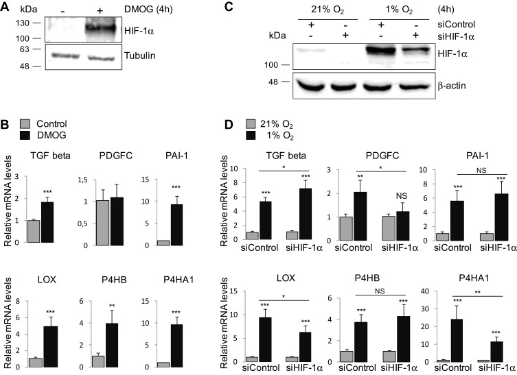
Hypoxia and its key mediators hypoxia inducible Factors (HIFs) are implicated in the development of liver diseases of diverse etiologies, often in interplay with inflammatory mediators. We investigated the interplay between hypoxia and proinflammatory mediators in the development of liver fibrosis, using human hepatocellular carcinoma Huh7 cells as a model. Treatment of Huh7 with DMOG or under hypoxia, induced HIF-1α protein levels and the expression of genes for pro-fibrotic (TGF-β1, PDGFC, PAI-1) and fibrosis (LOX, P4HA1, P4HB) markers. Knockdown of HIF-1α decreased the induction of PDGFC, LOX and P4HA1, showing the involvement of HIF-1 in their regulation. Interestingly, incubation of Huh7 cells under hypoxia did not cause activation of the NF-κΒ pathway. In contrast, inflammatory mediators such as tumor necrosis factor α (TNFα) and lipopolysaccharides (LPS) activated the NF-κΒ pathway, but failed to increase HIF-1α protein levels. Moreover, TNFα had a weaker effect than hypoxia on the induction or did not induce pro-fibrotic and fibrosis markers, respectively, while LPS enhanced only the hypoxic induction of P4HB. In conclusion, the above findings suggest that hypoxia and HIF-1 play an important role in the development of fibrosis in hepatocellular carcinoma, which appears to be independent of the activation of the NF-κΒ pathway.


 分享
分享

 求助内容:
求助内容: 应助结果提醒方式:
 扫码关注我们
