Tae-Hyun Choi, So-Hyun Kim, Cheul Kim, Yoon-Ah Kook, Brent E Larson, Nam-Ki Lee
{"title":"拔牙与非拔牙正畸治疗后最大闭唇力的变化。","authors":"Tae-Hyun Choi, So-Hyun Kim, Cheul Kim, Yoon-Ah Kook, Brent E Larson, Nam-Ki Lee","doi":"10.4041/kjod.2020.50.2.120","DOIUrl":null,"url":null,"abstract":"<p><strong>Objective: </strong>The aims of the present study were to evaluate the changes in the maximum lip-closing force (MLF) after orthodontic treatment with or without premolar extractions and verify the correlation of these changes with dentoskeletal changes.</p><p><strong>Methods: </strong>In total, 17 women who underwent nonextraction orthodontic treatment and 15 women who underwent orthodontic treatment with extraction of all four first premolars were included in this retrospective study. For all patients, lateral cephalograms and dental models were measured before (T0) and after (T1) treatment. In addition, MLF was measured at both time points using the Lip De Cum LDC-110R® device. Statistical analyses were performed to evaluate changes in clinical variables and MLF and their correlations.</p><p><strong>Results: </strong>Both groups showed similar skeletal patterns, although the extraction group showed greater proclination of the maxillary and mandibular incisors and lip protrusion compared to the nonextraction group at T0. MLF at T0 was comparable between the two groups. The reduction in the arch width and depth and incisor retroclination from T0 to T1 were more pronounced in the extraction group than in the nonextraction group. MLF in the extraction group significantly increased during the treatment period, and this increase was significantly greater than that in the nonextraction group. The increase in MLF was found to be correlated with the increase in the interincisal angle and decrease in the intermolar width, arch depth, and incisor-mandibular plane angle.</p><p><strong>Conclusions: </strong>This study suggests that MLF increases to a greater extent during extraction orthodontic treatment than during nonextraction orthodontic treatment.</p>","PeriodicalId":49934,"journal":{"name":"Korean Journal of Orthodontics","volume":null,"pages":null},"PeriodicalIF":1.9000,"publicationDate":"2020-03-01","publicationTypes":"Journal Article","fieldsOfStudy":null,"isOpenAccess":false,"openAccessPdf":"https://sci-hub-pdf.com/10.4041/kjod.2020.50.2.120","citationCount":"2","resultStr":"{\"title\":\"Changes in maximum lip-closing force after extraction and nonextraction orthodontic treatments.\",\"authors\":\"Tae-Hyun Choi, So-Hyun Kim, Cheul Kim, Yoon-Ah Kook, Brent E Larson, Nam-Ki Lee\",\"doi\":\"10.4041/kjod.2020.50.2.120\",\"DOIUrl\":null,\"url\":null,\"abstract\":\"<p><strong>Objective: </strong>The aims of the present study were to evaluate the changes in the maximum lip-closing force (MLF) after orthodontic treatment with or without premolar extractions and verify the correlation of these changes with dentoskeletal changes.</p><p><strong>Methods: </strong>In total, 17 women who underwent nonextraction orthodontic treatment and 15 women who underwent orthodontic treatment with extraction of all four first premolars were included in this retrospective study. For all patients, lateral cephalograms and dental models were measured before (T0) and after (T1) treatment. In addition, MLF was measured at both time points using the Lip De Cum LDC-110R® device. Statistical analyses were performed to evaluate changes in clinical variables and MLF and their correlations.</p><p><strong>Results: </strong>Both groups showed similar skeletal patterns, although the extraction group showed greater proclination of the maxillary and mandibular incisors and lip protrusion compared to the nonextraction group at T0. MLF at T0 was comparable between the two groups. The reduction in the arch width and depth and incisor retroclination from T0 to T1 were more pronounced in the extraction group than in the nonextraction group. MLF in the extraction group significantly increased during the treatment period, and this increase was significantly greater than that in the nonextraction group. The increase in MLF was found to be correlated with the increase in the interincisal angle and decrease in the intermolar width, arch depth, and incisor-mandibular plane angle.</p><p><strong>Conclusions: </strong>This study suggests that MLF increases to a greater extent during extraction orthodontic treatment than during nonextraction orthodontic treatment.</p>\",\"PeriodicalId\":49934,\"journal\":{\"name\":\"Korean Journal of Orthodontics\",\"volume\":null,\"pages\":null},\"PeriodicalIF\":1.9000,\"publicationDate\":\"2020-03-01\",\"publicationTypes\":\"Journal Article\",\"fieldsOfStudy\":null,\"isOpenAccess\":false,\"openAccessPdf\":\"https://sci-hub-pdf.com/10.4041/kjod.2020.50.2.120\",\"citationCount\":\"2\",\"resultStr\":null,\"platform\":\"Semanticscholar\",\"paperid\":null,\"PeriodicalName\":\"Korean Journal of Orthodontics\",\"FirstCategoryId\":\"3\",\"ListUrlMain\":\"https://doi.org/10.4041/kjod.2020.50.2.120\",\"RegionNum\":3,\"RegionCategory\":\"医学\",\"ArticlePicture\":[],\"TitleCN\":null,\"AbstractTextCN\":null,\"PMCID\":null,\"EPubDate\":\"2020/3/24 0:00:00\",\"PubModel\":\"Epub\",\"JCR\":\"Q1\",\"JCRName\":\"Dentistry\",\"Score\":null,\"Total\":0}","platform":"Semanticscholar","paperid":null,"PeriodicalName":"Korean Journal of Orthodontics","FirstCategoryId":"3","ListUrlMain":"https://doi.org/10.4041/kjod.2020.50.2.120","RegionNum":3,"RegionCategory":"医学","ArticlePicture":[],"TitleCN":null,"AbstractTextCN":null,"PMCID":null,"EPubDate":"2020/3/24 0:00:00","PubModel":"Epub","JCR":"Q1","JCRName":"Dentistry","Score":null,"Total":0}
引用次数: 2
摘要
目的:评价正畸治疗前后拔前磨牙和不拔前磨牙的最大闭唇力(MLF)的变化,并验证其与牙骨骼变化的相关性。方法:回顾性研究17例接受非拔牙正畸治疗的妇女和15例接受全部4颗第一前磨牙拔牙正畸治疗的妇女。所有患者在治疗前(T0)和治疗后(T1)分别测量侧位脑电图和牙模型。此外,使用Lip De Cum LDC-110R®装置在两个时间点测量MLF。对临床变量和MLF的变化及其相关性进行统计分析。结果:两组骨骼形态相似,但拔牙组在T0时上颌门牙较未拔牙组明显前倾,唇部较未拔牙组突出。两组在T0时的MLF具有可比性。拔牙组牙弓宽度、深度和切牙后倾从T0到T1的减少比未拔牙组更明显。拔牙组的MLF在治疗期间显著增加,且明显大于未拔牙组。MLF的增加与磨牙间角的增加、磨牙间宽度、牙弓深度和切下颌平面角的减小有关。结论:本研究提示拔牙正畸治疗中MLF的增加程度大于非拔牙正畸治疗。
Changes in maximum lip-closing force after extraction and nonextraction orthodontic treatments.
Objective: The aims of the present study were to evaluate the changes in the maximum lip-closing force (MLF) after orthodontic treatment with or without premolar extractions and verify the correlation of these changes with dentoskeletal changes.
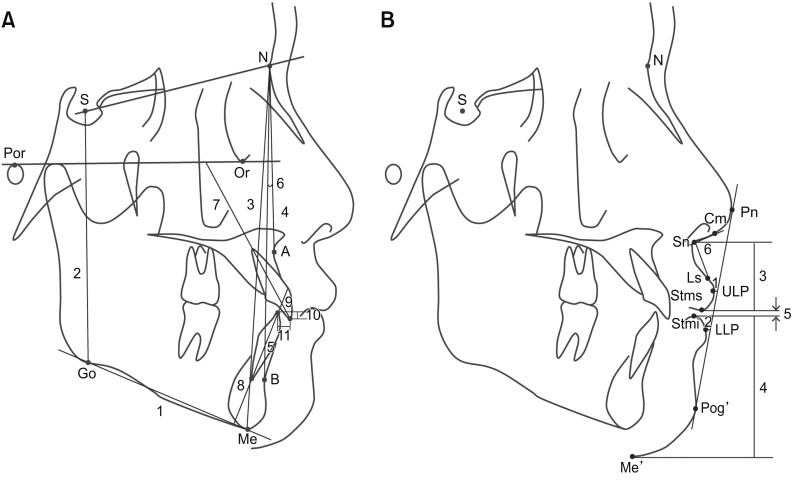
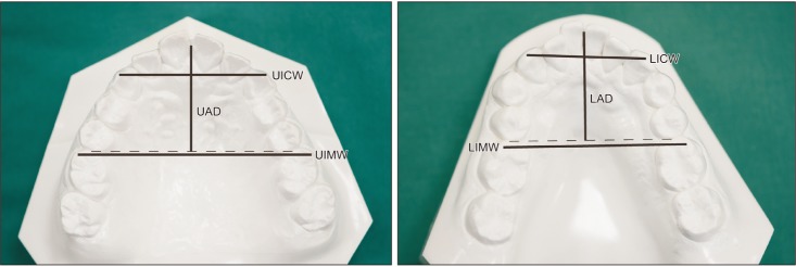
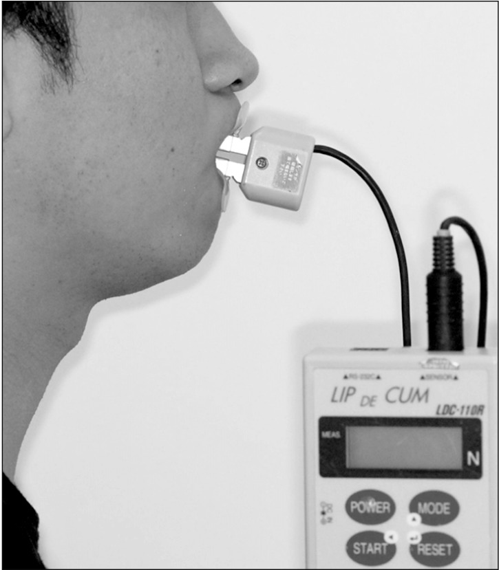
Methods: In total, 17 women who underwent nonextraction orthodontic treatment and 15 women who underwent orthodontic treatment with extraction of all four first premolars were included in this retrospective study. For all patients, lateral cephalograms and dental models were measured before (T0) and after (T1) treatment. In addition, MLF was measured at both time points using the Lip De Cum LDC-110R® device. Statistical analyses were performed to evaluate changes in clinical variables and MLF and their correlations.
Results: Both groups showed similar skeletal patterns, although the extraction group showed greater proclination of the maxillary and mandibular incisors and lip protrusion compared to the nonextraction group at T0. MLF at T0 was comparable between the two groups. The reduction in the arch width and depth and incisor retroclination from T0 to T1 were more pronounced in the extraction group than in the nonextraction group. MLF in the extraction group significantly increased during the treatment period, and this increase was significantly greater than that in the nonextraction group. The increase in MLF was found to be correlated with the increase in the interincisal angle and decrease in the intermolar width, arch depth, and incisor-mandibular plane angle.
Conclusions: This study suggests that MLF increases to a greater extent during extraction orthodontic treatment than during nonextraction orthodontic treatment.
期刊介绍:
The Korean Journal of Orthodontics (KJO) is an international, open access, peer reviewed journal published in January, March, May, July, September, and November each year. It was first launched in 1970 and, as the official scientific publication of Korean Association of Orthodontists, KJO aims to publish high quality clinical and scientific original research papers in all areas related to orthodontics and dentofacial orthopedics. Specifically, its interest focuses on evidence-based investigations of contemporary diagnostic procedures and treatment techniques, expanding to significant clinical reports of diverse treatment approaches.
The scope of KJO covers all areas of orthodontics and dentofacial orthopedics including successful diagnostic procedures and treatment planning, growth and development of the face and its clinical implications, appliance designs, biomechanics, TMJ disorders and adult treatment. Specifically, its latest interest focuses on skeletal anchorage devices, orthodontic appliance and biomaterials, 3 dimensional imaging techniques utilized for dentofacial diagnosis and treatment planning, and orthognathic surgery to correct skeletal disharmony in association of orthodontic treatment.


 分享
分享
 求助内容:
求助内容: 应助结果提醒方式:
 扫码关注我们
