Chun-Yao Chen, Kai-Chieh Hsu, Hsuan-Yu Yeh, Han-Chen Ho
{"title":"抗生素对小鼠结肠粘液层影响的可视化。","authors":"Chun-Yao Chen, Kai-Chieh Hsu, Hsuan-Yu Yeh, Han-Chen Ho","doi":"10.4103/tcmj.tcmj_70_19","DOIUrl":null,"url":null,"abstract":"<p><strong>Objective: </strong>Mucus provides a protective barrier separating sensitive epithelial surfaces from the outside world. The mouse colonic mucus is organized as a bacteria-free inner layer and a bacteria-colonized outer layer. Antibiotic treatments are known to disturb gut microbiota, but their effect on the mucosal barrier is rarely discussed. The aim was to evaluate and visualize the impact of antibiotics on the colonic mucus and the microbial community.</p><p><strong>Materials and methods: </strong>Two sets of experiments were conducted. In the antibiotic experiment, mice orally ingested both streptomycin and bacitracin for 7 days. In the recovery experiment, mice were allowed to recover for 7 days without antibiotics after having received the 7-day antibiotic treatment. Mouse colons were isolated and divided into proximal, middle, and distal parts. Specimens were examined under a transmission electron microscope to identify morphological changes. The gut microbial community was evaluated by analyzing 16S rDNA sequences isolated from the different parts of the mouse colon.</p><p><strong>Results: </strong>The antibiotic-treated mice were physiologically normal. However, a significantly increased inner mucus layer in the proximal and middle colon and a dramatic decrease in bacterial numbers in the outer mucus layers were observed. The 16S rDNA compositions showed a similarity in the dominant taxa among different colon sections. While control mice had a diverse microbiota, antibiotic treatments effectively eliminated most of the bacteria, such that the community was dominated by only one genus (<i>Turicibacter</i> or <i>Staphylococcus</i>). Furthermore, following antibiotic withdrawal in treated mice, the thickness of the inner mucus layer returned to control levels, and the microbial community regained a more complex structure, dominated by <i>Firmicutes</i>, <i>Bacteroidetes</i>, and <i>Proteobacteria</i>.</p><p><strong>Conclusions: </strong>Our results indicated that antibiotic treatments not only disturbed the microbiota but also altered the structure of the mucus layer. After the withdrawal of antibiotics, the mucus layer was quickly regenerated within days, probably in response to microbial growth. The recolonization by gut inhabitants with diverse ecological roles, such as mucin-degraders and fermenters indicate that the gut ecosystem is functionally sound and highly resilient.</p>","PeriodicalId":72593,"journal":{"name":"Ci ji yi xue za zhi = Tzu-chi medical journal","volume":"32 2","pages":"145-153"},"PeriodicalIF":0.0000,"publicationDate":"2019-08-20","publicationTypes":"Journal Article","fieldsOfStudy":null,"isOpenAccess":false,"openAccessPdf":"https://ftp.ncbi.nlm.nih.gov/pub/pmc/oa_pdf/da/0a/TCMJ-32-145.PMC7137371.pdf","citationCount":"0","resultStr":"{\"title\":\"Visualizing the effects of antibiotics on the mouse colonic mucus layer.\",\"authors\":\"Chun-Yao Chen, Kai-Chieh Hsu, Hsuan-Yu Yeh, Han-Chen Ho\",\"doi\":\"10.4103/tcmj.tcmj_70_19\",\"DOIUrl\":null,\"url\":null,\"abstract\":\"<p><strong>Objective: </strong>Mucus provides a protective barrier separating sensitive epithelial surfaces from the outside world. The mouse colonic mucus is organized as a bacteria-free inner layer and a bacteria-colonized outer layer. Antibiotic treatments are known to disturb gut microbiota, but their effect on the mucosal barrier is rarely discussed. The aim was to evaluate and visualize the impact of antibiotics on the colonic mucus and the microbial community.</p><p><strong>Materials and methods: </strong>Two sets of experiments were conducted. In the antibiotic experiment, mice orally ingested both streptomycin and bacitracin for 7 days. In the recovery experiment, mice were allowed to recover for 7 days without antibiotics after having received the 7-day antibiotic treatment. Mouse colons were isolated and divided into proximal, middle, and distal parts. Specimens were examined under a transmission electron microscope to identify morphological changes. The gut microbial community was evaluated by analyzing 16S rDNA sequences isolated from the different parts of the mouse colon.</p><p><strong>Results: </strong>The antibiotic-treated mice were physiologically normal. However, a significantly increased inner mucus layer in the proximal and middle colon and a dramatic decrease in bacterial numbers in the outer mucus layers were observed. The 16S rDNA compositions showed a similarity in the dominant taxa among different colon sections. While control mice had a diverse microbiota, antibiotic treatments effectively eliminated most of the bacteria, such that the community was dominated by only one genus (<i>Turicibacter</i> or <i>Staphylococcus</i>). Furthermore, following antibiotic withdrawal in treated mice, the thickness of the inner mucus layer returned to control levels, and the microbial community regained a more complex structure, dominated by <i>Firmicutes</i>, <i>Bacteroidetes</i>, and <i>Proteobacteria</i>.</p><p><strong>Conclusions: </strong>Our results indicated that antibiotic treatments not only disturbed the microbiota but also altered the structure of the mucus layer. After the withdrawal of antibiotics, the mucus layer was quickly regenerated within days, probably in response to microbial growth. The recolonization by gut inhabitants with diverse ecological roles, such as mucin-degraders and fermenters indicate that the gut ecosystem is functionally sound and highly resilient.</p>\",\"PeriodicalId\":72593,\"journal\":{\"name\":\"Ci ji yi xue za zhi = Tzu-chi medical journal\",\"volume\":\"32 2\",\"pages\":\"145-153\"},\"PeriodicalIF\":0.0000,\"publicationDate\":\"2019-08-20\",\"publicationTypes\":\"Journal Article\",\"fieldsOfStudy\":null,\"isOpenAccess\":false,\"openAccessPdf\":\"https://ftp.ncbi.nlm.nih.gov/pub/pmc/oa_pdf/da/0a/TCMJ-32-145.PMC7137371.pdf\",\"citationCount\":\"0\",\"resultStr\":null,\"platform\":\"Semanticscholar\",\"paperid\":null,\"PeriodicalName\":\"Ci ji yi xue za zhi = Tzu-chi medical journal\",\"FirstCategoryId\":\"1085\",\"ListUrlMain\":\"https://doi.org/10.4103/tcmj.tcmj_70_19\",\"RegionNum\":0,\"RegionCategory\":null,\"ArticlePicture\":[],\"TitleCN\":null,\"AbstractTextCN\":null,\"PMCID\":null,\"EPubDate\":\"2020/4/1 0:00:00\",\"PubModel\":\"eCollection\",\"JCR\":\"\",\"JCRName\":\"\",\"Score\":null,\"Total\":0}","platform":"Semanticscholar","paperid":null,"PeriodicalName":"Ci ji yi xue za zhi = Tzu-chi medical journal","FirstCategoryId":"1085","ListUrlMain":"https://doi.org/10.4103/tcmj.tcmj_70_19","RegionNum":0,"RegionCategory":null,"ArticlePicture":[],"TitleCN":null,"AbstractTextCN":null,"PMCID":null,"EPubDate":"2020/4/1 0:00:00","PubModel":"eCollection","JCR":"","JCRName":"","Score":null,"Total":0}
Visualizing the effects of antibiotics on the mouse colonic mucus layer.
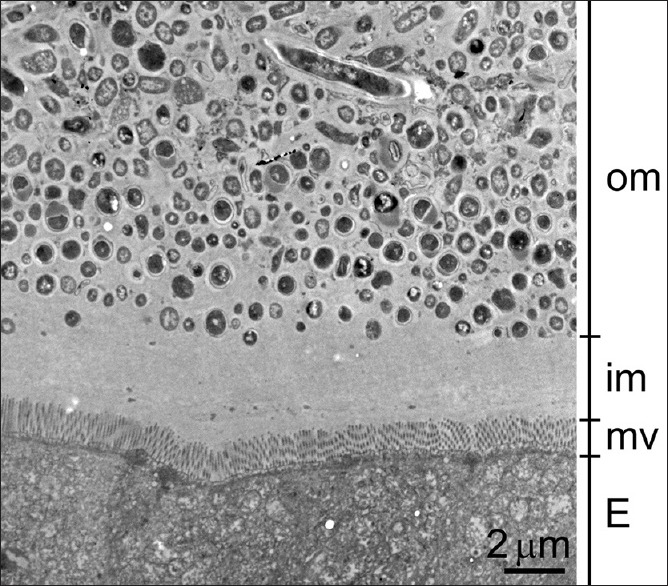
Objective: Mucus provides a protective barrier separating sensitive epithelial surfaces from the outside world. The mouse colonic mucus is organized as a bacteria-free inner layer and a bacteria-colonized outer layer. Antibiotic treatments are known to disturb gut microbiota, but their effect on the mucosal barrier is rarely discussed. The aim was to evaluate and visualize the impact of antibiotics on the colonic mucus and the microbial community.
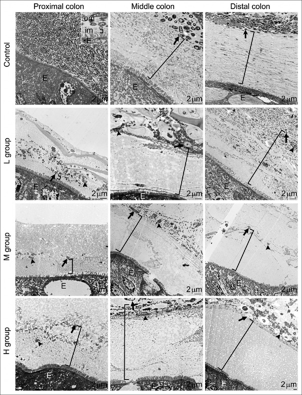
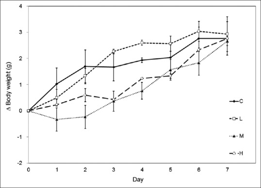
Materials and methods: Two sets of experiments were conducted. In the antibiotic experiment, mice orally ingested both streptomycin and bacitracin for 7 days. In the recovery experiment, mice were allowed to recover for 7 days without antibiotics after having received the 7-day antibiotic treatment. Mouse colons were isolated and divided into proximal, middle, and distal parts. Specimens were examined under a transmission electron microscope to identify morphological changes. The gut microbial community was evaluated by analyzing 16S rDNA sequences isolated from the different parts of the mouse colon.
Results: The antibiotic-treated mice were physiologically normal. However, a significantly increased inner mucus layer in the proximal and middle colon and a dramatic decrease in bacterial numbers in the outer mucus layers were observed. The 16S rDNA compositions showed a similarity in the dominant taxa among different colon sections. While control mice had a diverse microbiota, antibiotic treatments effectively eliminated most of the bacteria, such that the community was dominated by only one genus (Turicibacter or Staphylococcus). Furthermore, following antibiotic withdrawal in treated mice, the thickness of the inner mucus layer returned to control levels, and the microbial community regained a more complex structure, dominated by Firmicutes, Bacteroidetes, and Proteobacteria.
Conclusions: Our results indicated that antibiotic treatments not only disturbed the microbiota but also altered the structure of the mucus layer. After the withdrawal of antibiotics, the mucus layer was quickly regenerated within days, probably in response to microbial growth. The recolonization by gut inhabitants with diverse ecological roles, such as mucin-degraders and fermenters indicate that the gut ecosystem is functionally sound and highly resilient.


 分享
分享

 求助内容:
求助内容: 应助结果提醒方式:
 扫码关注我们
