{"title":"胆绿素还原酶-A 缺乏会使胆绿素结合色蛋白变亮和敏感。","authors":"Kenju Kobachi, Sota Kuno, Shinya Sato, Kenta Sumiyama, Michiyuki Matsuda, Kenta Terai","doi":"10.1247/csf.20010","DOIUrl":null,"url":null,"abstract":"<p><p>Tissue absorbance, light scattering, and autofluorescence are significantly lower in the near-infrared (NIR) range than in the visible range. Because of these advantages, NIR fluorescent proteins (FPs) are in high demand for in vivo imaging. Nevertheless, application of NIR FPs such as iRFP is still limited due to their dimness in mammalian cells. In contrast to GFP and its variants, iRFP requires biliverdin (BV) as a chromophore. The dimness of iRFP is at least partly due to rapid reduction of BV by biliverdin reductase-A (BLVRA). Here, we established biliverdin reductase-a knockout (Blvra<sup>-/-</sup>) mice to increase the intracellular BV concentration and, thereby, to enhance iRFP fluorescence intensity. As anticipated, iRFP fluorescence intensity was significantly increased in all examined tissues of Blvra<sup>-/-</sup> mice. Similarly, the genetically encoded calcium indicator NIR-GECO1, which is engineered based on another NIR FP, mIFP, exhibited a marked increase in fluorescence intensity in mouse embryonic fibroblasts derived from Blvra<sup>-/-</sup> mice. We expanded this approach to an NIR light-sensing optogenetic tool, the BphP1-PpsR2 system, which also requires BV as a chromophore. Again, deletion of the Blvra gene markedly enhanced the light response in HeLa cells. These results indicate that the Blvra<sup>-/-</sup> mouse is a versatile tool for the in vivo application of NIR FPs and NIR light-sensing optogenetic tools.Key words: in vivo imaging, near-infrared fluorescent protein, biliverdin, biliverdin reductase, optogenetic tool.</p>","PeriodicalId":9927,"journal":{"name":"Cell structure and function","volume":"45 2","pages":"131-141"},"PeriodicalIF":2.2000,"publicationDate":"2020-08-21","publicationTypes":"Journal Article","fieldsOfStudy":null,"isOpenAccess":false,"openAccessPdf":"https://www.ncbi.nlm.nih.gov/pmc/articles/PMC10511041/pdf/","citationCount":"11","resultStr":"{\"title\":\"Biliverdin Reductase-A Deficiency Brighten and Sensitize Biliverdin-binding Chromoproteins.\",\"authors\":\"Kenju Kobachi, Sota Kuno, Shinya Sato, Kenta Sumiyama, Michiyuki Matsuda, Kenta Terai\",\"doi\":\"10.1247/csf.20010\",\"DOIUrl\":null,\"url\":null,\"abstract\":\"<p><p>Tissue absorbance, light scattering, and autofluorescence are significantly lower in the near-infrared (NIR) range than in the visible range. Because of these advantages, NIR fluorescent proteins (FPs) are in high demand for in vivo imaging. Nevertheless, application of NIR FPs such as iRFP is still limited due to their dimness in mammalian cells. In contrast to GFP and its variants, iRFP requires biliverdin (BV) as a chromophore. The dimness of iRFP is at least partly due to rapid reduction of BV by biliverdin reductase-A (BLVRA). Here, we established biliverdin reductase-a knockout (Blvra<sup>-/-</sup>) mice to increase the intracellular BV concentration and, thereby, to enhance iRFP fluorescence intensity. As anticipated, iRFP fluorescence intensity was significantly increased in all examined tissues of Blvra<sup>-/-</sup> mice. Similarly, the genetically encoded calcium indicator NIR-GECO1, which is engineered based on another NIR FP, mIFP, exhibited a marked increase in fluorescence intensity in mouse embryonic fibroblasts derived from Blvra<sup>-/-</sup> mice. We expanded this approach to an NIR light-sensing optogenetic tool, the BphP1-PpsR2 system, which also requires BV as a chromophore. Again, deletion of the Blvra gene markedly enhanced the light response in HeLa cells. These results indicate that the Blvra<sup>-/-</sup> mouse is a versatile tool for the in vivo application of NIR FPs and NIR light-sensing optogenetic tools.Key words: in vivo imaging, near-infrared fluorescent protein, biliverdin, biliverdin reductase, optogenetic tool.</p>\",\"PeriodicalId\":9927,\"journal\":{\"name\":\"Cell structure and function\",\"volume\":\"45 2\",\"pages\":\"131-141\"},\"PeriodicalIF\":2.2000,\"publicationDate\":\"2020-08-21\",\"publicationTypes\":\"Journal Article\",\"fieldsOfStudy\":null,\"isOpenAccess\":false,\"openAccessPdf\":\"https://www.ncbi.nlm.nih.gov/pmc/articles/PMC10511041/pdf/\",\"citationCount\":\"11\",\"resultStr\":null,\"platform\":\"Semanticscholar\",\"paperid\":null,\"PeriodicalName\":\"Cell structure and function\",\"FirstCategoryId\":\"99\",\"ListUrlMain\":\"https://doi.org/10.1247/csf.20010\",\"RegionNum\":4,\"RegionCategory\":\"生物学\",\"ArticlePicture\":[],\"TitleCN\":null,\"AbstractTextCN\":null,\"PMCID\":null,\"EPubDate\":\"2020/6/25 0:00:00\",\"PubModel\":\"Epub\",\"JCR\":\"Q4\",\"JCRName\":\"CELL BIOLOGY\",\"Score\":null,\"Total\":0}","platform":"Semanticscholar","paperid":null,"PeriodicalName":"Cell structure and function","FirstCategoryId":"99","ListUrlMain":"https://doi.org/10.1247/csf.20010","RegionNum":4,"RegionCategory":"生物学","ArticlePicture":[],"TitleCN":null,"AbstractTextCN":null,"PMCID":null,"EPubDate":"2020/6/25 0:00:00","PubModel":"Epub","JCR":"Q4","JCRName":"CELL BIOLOGY","Score":null,"Total":0}
引用次数: 11
摘要
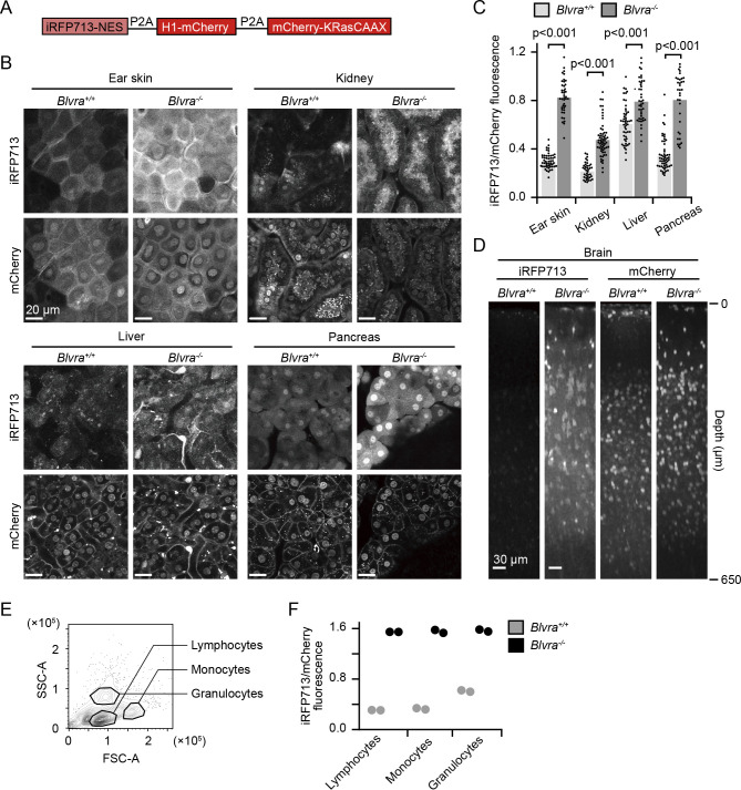
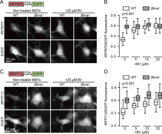
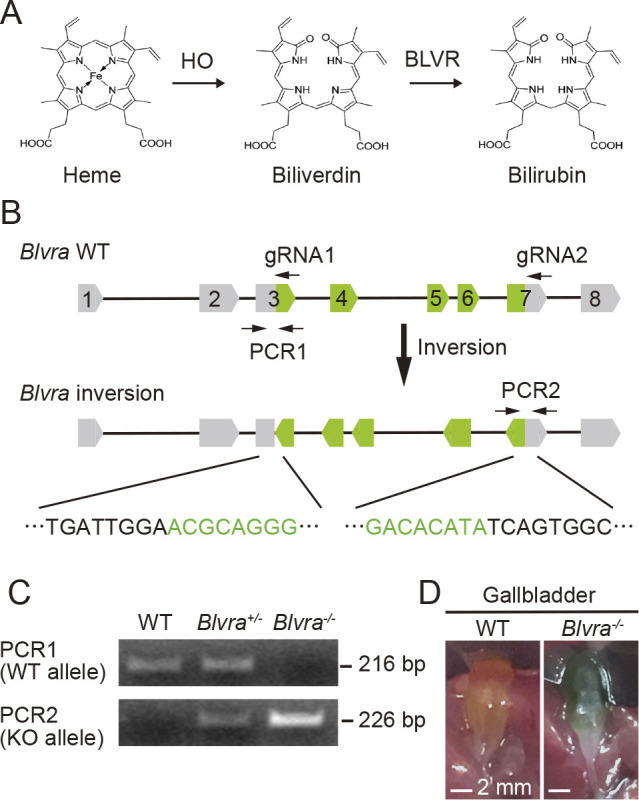
组织吸光度、光散射和自身荧光在近红外(NIR)范围内明显低于可见光范围。由于这些优点,近红外荧光蛋白(FPs)在体内成像中有很高的需求。然而,近红外荧光蛋白(如iRFP)的应用仍然受到限制,因为它们在哺乳动物细胞中较暗。与GFP及其变体不同,iRFP需要胆绿素(BV)作为发色团。iRFP的模糊至少部分是由于胆绿素还原酶- a (BLVRA)快速还原BV。在这里,我们建立了胆绿素还原酶-a敲除(Blvra-/-)小鼠,以增加细胞内BV浓度,从而增强iRFP荧光强度。正如预期的那样,iRFP荧光强度在Blvra-/-小鼠的所有检查组织中显著增加。同样,基因编码的钙指示剂NIR- geco1,基于另一种近红外FP, mIFP,在来源于Blvra-/-小鼠的小鼠胚胎成纤维细胞中显示出显著的荧光强度增加。我们将这种方法扩展到近红外光传感光遗传工具BphP1-PpsR2系统,该系统也需要BV作为发色团。再一次,Blvra基因的缺失显著增强了HeLa细胞的光反应。这些结果表明,Blvra-/-小鼠是近红外FPs和近红外光敏光遗传工具在体内应用的多功能工具。关键词:体内成像,近红外荧光蛋白,胆绿素,胆绿素还原酶,光遗传工具
Biliverdin Reductase-A Deficiency Brighten and Sensitize Biliverdin-binding Chromoproteins.
Tissue absorbance, light scattering, and autofluorescence are significantly lower in the near-infrared (NIR) range than in the visible range. Because of these advantages, NIR fluorescent proteins (FPs) are in high demand for in vivo imaging. Nevertheless, application of NIR FPs such as iRFP is still limited due to their dimness in mammalian cells. In contrast to GFP and its variants, iRFP requires biliverdin (BV) as a chromophore. The dimness of iRFP is at least partly due to rapid reduction of BV by biliverdin reductase-A (BLVRA). Here, we established biliverdin reductase-a knockout (Blvra-/-) mice to increase the intracellular BV concentration and, thereby, to enhance iRFP fluorescence intensity. As anticipated, iRFP fluorescence intensity was significantly increased in all examined tissues of Blvra-/- mice. Similarly, the genetically encoded calcium indicator NIR-GECO1, which is engineered based on another NIR FP, mIFP, exhibited a marked increase in fluorescence intensity in mouse embryonic fibroblasts derived from Blvra-/- mice. We expanded this approach to an NIR light-sensing optogenetic tool, the BphP1-PpsR2 system, which also requires BV as a chromophore. Again, deletion of the Blvra gene markedly enhanced the light response in HeLa cells. These results indicate that the Blvra-/- mouse is a versatile tool for the in vivo application of NIR FPs and NIR light-sensing optogenetic tools.Key words: in vivo imaging, near-infrared fluorescent protein, biliverdin, biliverdin reductase, optogenetic tool.
期刊介绍:
Cell Structure and Function is a fully peer-reviewed, fully Open Access journal. As the official English-language journal of the Japan Society for Cell Biology, it is published continuously online and biannually in print.
Cell Structure and Function publishes important, original contributions in all areas of molecular and cell biology. The journal welcomes the submission of manuscripts on research areas such as the cell nucleus, chromosomes, and gene expression; the cytoskeleton and cell motility; cell adhesion and the extracellular matrix; cell growth, differentiation and death; signal transduction; the protein life cycle; membrane traffic; and organelles.


 分享
分享
 求助内容:
求助内容: 应助结果提醒方式:
 扫码关注我们
