Kimberly C. Grasty, Stephen D. Weeks , Patrick J. Loll
{"title":"人类Josephin-2活性和调控的结构见解","authors":"Kimberly C. Grasty, Stephen D. Weeks , Patrick J. Loll","doi":"10.1016/j.yjsbx.2019.100011","DOIUrl":null,"url":null,"abstract":"<div><p>The MJD family of human deubiquitinating enzymes contains four members: Ataxin-3, the ataxin-3-like protein (AT3L), Josephin-1, and Josephin-2. All share a conserved catalytic unit known as the Josephin domain. Ataxin-3 and AT3L also contain extensive regulatory regions that modulate their functions, whereas Josephins-1 and -2 are substantially smaller, containing only the Josephin domain. To gain insight into how these minimal Josephins differ from their larger relatives, we determined the 2.3 Å X-ray crystal structure of human Josephin-2 and probed the enzyme’s substrate specificity. Several large disordered loops are seen in the structure, suggesting a highly dynamic enzyme. Josephin-2 lacks several allosteric sites found in ataxin-3, but its structure suggests potential regulation via ubiquitination of a loop adjoining the active site. The enzyme preferentially recognizes substrates containing K11, K48, and K63 linkages, pointing toward a possible role in maintenance of protein quality control.</p></div>","PeriodicalId":17238,"journal":{"name":"Journal of Structural Biology: X","volume":"3 ","pages":"Article 100011"},"PeriodicalIF":5.1000,"publicationDate":"2019-07-01","publicationTypes":"Journal Article","fieldsOfStudy":null,"isOpenAccess":false,"openAccessPdf":"https://sci-hub-pdf.com/10.1016/j.yjsbx.2019.100011","citationCount":"10","resultStr":"{\"title\":\"Structural insights into the activity and regulation of human Josephin-2\",\"authors\":\"Kimberly C. Grasty, Stephen D. Weeks , Patrick J. Loll\",\"doi\":\"10.1016/j.yjsbx.2019.100011\",\"DOIUrl\":null,\"url\":null,\"abstract\":\"<div><p>The MJD family of human deubiquitinating enzymes contains four members: Ataxin-3, the ataxin-3-like protein (AT3L), Josephin-1, and Josephin-2. All share a conserved catalytic unit known as the Josephin domain. Ataxin-3 and AT3L also contain extensive regulatory regions that modulate their functions, whereas Josephins-1 and -2 are substantially smaller, containing only the Josephin domain. To gain insight into how these minimal Josephins differ from their larger relatives, we determined the 2.3 Å X-ray crystal structure of human Josephin-2 and probed the enzyme’s substrate specificity. Several large disordered loops are seen in the structure, suggesting a highly dynamic enzyme. Josephin-2 lacks several allosteric sites found in ataxin-3, but its structure suggests potential regulation via ubiquitination of a loop adjoining the active site. The enzyme preferentially recognizes substrates containing K11, K48, and K63 linkages, pointing toward a possible role in maintenance of protein quality control.</p></div>\",\"PeriodicalId\":17238,\"journal\":{\"name\":\"Journal of Structural Biology: X\",\"volume\":\"3 \",\"pages\":\"Article 100011\"},\"PeriodicalIF\":5.1000,\"publicationDate\":\"2019-07-01\",\"publicationTypes\":\"Journal Article\",\"fieldsOfStudy\":null,\"isOpenAccess\":false,\"openAccessPdf\":\"https://sci-hub-pdf.com/10.1016/j.yjsbx.2019.100011\",\"citationCount\":\"10\",\"resultStr\":null,\"platform\":\"Semanticscholar\",\"paperid\":null,\"PeriodicalName\":\"Journal of Structural Biology: X\",\"FirstCategoryId\":\"1085\",\"ListUrlMain\":\"https://www.sciencedirect.com/science/article/pii/S2590152419300091\",\"RegionNum\":0,\"RegionCategory\":null,\"ArticlePicture\":[],\"TitleCN\":null,\"AbstractTextCN\":null,\"PMCID\":null,\"EPubDate\":\"2019/8/21 0:00:00\",\"PubModel\":\"Epub\",\"JCR\":\"Q2\",\"JCRName\":\"BIOCHEMISTRY & MOLECULAR BIOLOGY\",\"Score\":null,\"Total\":0}","platform":"Semanticscholar","paperid":null,"PeriodicalName":"Journal of Structural Biology: X","FirstCategoryId":"1085","ListUrlMain":"https://www.sciencedirect.com/science/article/pii/S2590152419300091","RegionNum":0,"RegionCategory":null,"ArticlePicture":[],"TitleCN":null,"AbstractTextCN":null,"PMCID":null,"EPubDate":"2019/8/21 0:00:00","PubModel":"Epub","JCR":"Q2","JCRName":"BIOCHEMISTRY & MOLECULAR BIOLOGY","Score":null,"Total":0}
引用次数: 10
摘要
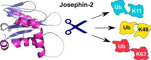
人去泛素化酶MJD家族包含四个成员:Ataxin-3、Ataxin-3样蛋白(AT3L)、Josephin-1和Josephin-2。它们都有一个保守的催化单元,称为约瑟夫结构域。Ataxin-3和AT3L也包含广泛的调节其功能的调控区域,而Josephin -1和-2则要小得多,只包含Josephin结构域。为了深入了解这些最小的josephin与较大的josephin有何不同,我们确定了人类Josephin-2的2.3 Å x射线晶体结构,并探测了该酶的底物特异性。在结构中可以看到几个大的无序环,表明这是一种高度动态的酶。Josephin-2缺乏在ataxin-3中发现的几个变构位点,但其结构表明可能通过邻近活性位点的环的泛素化进行调节。该酶优先识别含有K11、K48和K63键的底物,这表明它可能在维持蛋白质质量控制中起作用。
Structural insights into the activity and regulation of human Josephin-2
The MJD family of human deubiquitinating enzymes contains four members: Ataxin-3, the ataxin-3-like protein (AT3L), Josephin-1, and Josephin-2. All share a conserved catalytic unit known as the Josephin domain. Ataxin-3 and AT3L also contain extensive regulatory regions that modulate their functions, whereas Josephins-1 and -2 are substantially smaller, containing only the Josephin domain. To gain insight into how these minimal Josephins differ from their larger relatives, we determined the 2.3 Å X-ray crystal structure of human Josephin-2 and probed the enzyme’s substrate specificity. Several large disordered loops are seen in the structure, suggesting a highly dynamic enzyme. Josephin-2 lacks several allosteric sites found in ataxin-3, but its structure suggests potential regulation via ubiquitination of a loop adjoining the active site. The enzyme preferentially recognizes substrates containing K11, K48, and K63 linkages, pointing toward a possible role in maintenance of protein quality control.


 分享
分享
 求助内容:
求助内容: 应助结果提醒方式:
 扫码关注我们
