Roland Plesker, Kernt Köhler, Susanne von Gerlach, Klaus Boller, Markus Vogt, Inke S Feder
{"title":"非洲绿猴(Chlorocebus aethiops)的反应性间皮瘤增生模拟间皮瘤。","authors":"Roland Plesker, Kernt Köhler, Susanne von Gerlach, Klaus Boller, Markus Vogt, Inke S Feder","doi":"10.5194/pb-7-5-2020","DOIUrl":null,"url":null,"abstract":"<p><p>A spontaneous reactive mesothelial hyperplasia occurred in a female, 15.7-year-old African green monkey (grivet; <i>Chlorocebus aethiops</i>). At necropsy, massive effusions were found in the abdomen, the thorax, and the pericardium. Additionally, multiple small, beige-gray nodules were detected on the serosal surfaces of the abdominal organs. Histopathologically, the mesothelial cells resembled the epithelioid subtype of a mesothelioma, but no infiltrative or invasive growth could be demonstrated. The mesothelial cells on the thoracis, liver, and intestinal serosa were accompanied by chronic serositis. Mesothelial cells expressed cytokeratin, vimentin, calretinin, desmin, Wilms Tumor 1 (WT-1) protein, and epithelial membrane antigen (EMA). Cells were negative for carcinoembryonic antigen (CEA), cluster of differentiation 15 (CD15), and podoplanin. Ultrastructurally, cells revealed a moderate amount of microvilli of medium length, perinuclear tonofilament bundles, and long desmosomes. In fluorescence in situ hybridization (FISH) for the detection of characteristic gene loss (p16; CDKN2A), NF2, and MTAP, no deletions were detected. No asbestos fibers and no presence of Simian virus 40 antigen (SV40) could be demonstrated.</p>","PeriodicalId":37245,"journal":{"name":"Primate Biology","volume":"7 1","pages":"5-12"},"PeriodicalIF":0.0000,"publicationDate":"2020-06-15","publicationTypes":"Journal Article","fieldsOfStudy":null,"isOpenAccess":false,"openAccessPdf":"https://www.ncbi.nlm.nih.gov/pmc/articles/PMC7399384/pdf/","citationCount":"1","resultStr":"{\"title\":\"Reactive mesothelial hyperplasia mimicking mesothelioma in an African green monkey (<i>Chlorocebus aethiops</i>).\",\"authors\":\"Roland Plesker, Kernt Köhler, Susanne von Gerlach, Klaus Boller, Markus Vogt, Inke S Feder\",\"doi\":\"10.5194/pb-7-5-2020\",\"DOIUrl\":null,\"url\":null,\"abstract\":\"<p><p>A spontaneous reactive mesothelial hyperplasia occurred in a female, 15.7-year-old African green monkey (grivet; <i>Chlorocebus aethiops</i>). At necropsy, massive effusions were found in the abdomen, the thorax, and the pericardium. Additionally, multiple small, beige-gray nodules were detected on the serosal surfaces of the abdominal organs. Histopathologically, the mesothelial cells resembled the epithelioid subtype of a mesothelioma, but no infiltrative or invasive growth could be demonstrated. The mesothelial cells on the thoracis, liver, and intestinal serosa were accompanied by chronic serositis. Mesothelial cells expressed cytokeratin, vimentin, calretinin, desmin, Wilms Tumor 1 (WT-1) protein, and epithelial membrane antigen (EMA). Cells were negative for carcinoembryonic antigen (CEA), cluster of differentiation 15 (CD15), and podoplanin. Ultrastructurally, cells revealed a moderate amount of microvilli of medium length, perinuclear tonofilament bundles, and long desmosomes. In fluorescence in situ hybridization (FISH) for the detection of characteristic gene loss (p16; CDKN2A), NF2, and MTAP, no deletions were detected. No asbestos fibers and no presence of Simian virus 40 antigen (SV40) could be demonstrated.</p>\",\"PeriodicalId\":37245,\"journal\":{\"name\":\"Primate Biology\",\"volume\":\"7 1\",\"pages\":\"5-12\"},\"PeriodicalIF\":0.0000,\"publicationDate\":\"2020-06-15\",\"publicationTypes\":\"Journal Article\",\"fieldsOfStudy\":null,\"isOpenAccess\":false,\"openAccessPdf\":\"https://www.ncbi.nlm.nih.gov/pmc/articles/PMC7399384/pdf/\",\"citationCount\":\"1\",\"resultStr\":null,\"platform\":\"Semanticscholar\",\"paperid\":null,\"PeriodicalName\":\"Primate Biology\",\"FirstCategoryId\":\"1085\",\"ListUrlMain\":\"https://doi.org/10.5194/pb-7-5-2020\",\"RegionNum\":0,\"RegionCategory\":null,\"ArticlePicture\":[],\"TitleCN\":null,\"AbstractTextCN\":null,\"PMCID\":null,\"EPubDate\":\"2020/1/1 0:00:00\",\"PubModel\":\"eCollection\",\"JCR\":\"Q2\",\"JCRName\":\"Agricultural and Biological Sciences\",\"Score\":null,\"Total\":0}","platform":"Semanticscholar","paperid":null,"PeriodicalName":"Primate Biology","FirstCategoryId":"1085","ListUrlMain":"https://doi.org/10.5194/pb-7-5-2020","RegionNum":0,"RegionCategory":null,"ArticlePicture":[],"TitleCN":null,"AbstractTextCN":null,"PMCID":null,"EPubDate":"2020/1/1 0:00:00","PubModel":"eCollection","JCR":"Q2","JCRName":"Agricultural and Biological Sciences","Score":null,"Total":0}
Reactive mesothelial hyperplasia mimicking mesothelioma in an African green monkey (Chlorocebus aethiops).
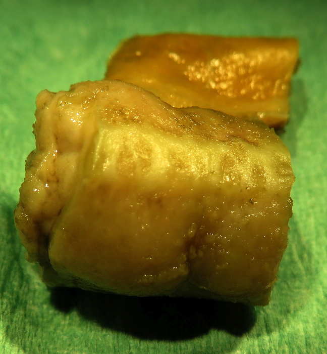
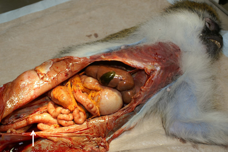
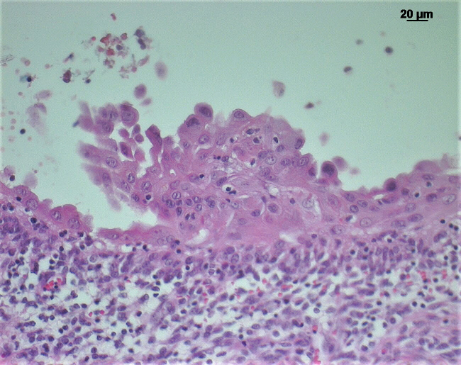
A spontaneous reactive mesothelial hyperplasia occurred in a female, 15.7-year-old African green monkey (grivet; Chlorocebus aethiops). At necropsy, massive effusions were found in the abdomen, the thorax, and the pericardium. Additionally, multiple small, beige-gray nodules were detected on the serosal surfaces of the abdominal organs. Histopathologically, the mesothelial cells resembled the epithelioid subtype of a mesothelioma, but no infiltrative or invasive growth could be demonstrated. The mesothelial cells on the thoracis, liver, and intestinal serosa were accompanied by chronic serositis. Mesothelial cells expressed cytokeratin, vimentin, calretinin, desmin, Wilms Tumor 1 (WT-1) protein, and epithelial membrane antigen (EMA). Cells were negative for carcinoembryonic antigen (CEA), cluster of differentiation 15 (CD15), and podoplanin. Ultrastructurally, cells revealed a moderate amount of microvilli of medium length, perinuclear tonofilament bundles, and long desmosomes. In fluorescence in situ hybridization (FISH) for the detection of characteristic gene loss (p16; CDKN2A), NF2, and MTAP, no deletions were detected. No asbestos fibers and no presence of Simian virus 40 antigen (SV40) could be demonstrated.


 分享
分享
 求助内容:
求助内容: 应助结果提醒方式:
 扫码关注我们
