Ralf P Friedrich, Eveline Schreiber, Rainer Tietze, Hai Yang, Christian Pilarsky, Christoph Alexiou
{"title":"细胞内无标记氧化铁纳米颗粒的全息层析显微镜定量和定位。","authors":"Ralf P Friedrich, Eveline Schreiber, Rainer Tietze, Hai Yang, Christian Pilarsky, Christoph Alexiou","doi":"10.2147/NSA.S282204","DOIUrl":null,"url":null,"abstract":"<p><strong>Background: </strong>The limitations of optical microscopy to determine the cellular localization of label-free nanoparticles prevent a solid prediction of the cellular effect of particles intended for medical applications. To avoid the strong physicochemical changes associated with fluorescent labelling, which often result in differences in cellular uptake, efficiency and toxicity of particles, novel detection techniques are required.</p><p><strong>Methods: </strong>In the present study, we determined the intracellular content of unlabeled SPIONs by analyzing refractive index (RI)-based images from holotomographic three-dimensional (3D) microscopy and side scatter data measured by flow cytometry. The results were compared with the actual cellular SPION amount as quantified by atomic emission spectroscopy (AES).</p><p><strong>Results: </strong>Live cell imaging by 3D holotomographic microscopy demonstrated cell-specific differences in intracellular nanoparticle uptake in different pancreatic cell lines. Thus, treatment of PANC-1<sup>SMAD4 (1-4)</sup> and PANC-1<sup>SMAD4 (2-6)</sup> with SPIONs resulted in a significant increase in number of areas with higher RI, whereas in PANC-1, SUIT-2 and PaCa DD183, only a minimal increase of spots with high RI was observed. The increase in areas with high RI was in accordance with the SPION content determined by quantitative iron measurements using AES. In contrast, determination of the SPION amount by flow cytometry was strongly cell type-dependent and did not allow the discrimination between intracellular and membrane-bound SPIONs. However, flow cytometry is a very rapid and reliable method to assess the cellular toxicity and allows an estimation of the cell-associated SPION content.</p><p><strong>Conclusion: </strong>Holotomographic 3D microscopy is a useful method to distinguish between intracellular and membrane-associated particles. Thus, it provides a valuable tool for scientists to evaluate the cellular localization and the particle load, which facilitates prediction of potential toxicity and efficiency of nanoparticles for medical applications.</p>","PeriodicalId":18881,"journal":{"name":"Nanotechnology, Science and Applications","volume":"13 ","pages":"119-130"},"PeriodicalIF":2.4000,"publicationDate":"2020-12-09","publicationTypes":"Journal Article","fieldsOfStudy":null,"isOpenAccess":false,"openAccessPdf":"https://sci-hub-pdf.com/10.2147/NSA.S282204","citationCount":"7","resultStr":"{\"title\":\"Intracellular Quantification and Localization of Label-Free Iron Oxide Nanoparticles by Holotomographic Microscopy.\",\"authors\":\"Ralf P Friedrich, Eveline Schreiber, Rainer Tietze, Hai Yang, Christian Pilarsky, Christoph Alexiou\",\"doi\":\"10.2147/NSA.S282204\",\"DOIUrl\":null,\"url\":null,\"abstract\":\"<p><strong>Background: </strong>The limitations of optical microscopy to determine the cellular localization of label-free nanoparticles prevent a solid prediction of the cellular effect of particles intended for medical applications. To avoid the strong physicochemical changes associated with fluorescent labelling, which often result in differences in cellular uptake, efficiency and toxicity of particles, novel detection techniques are required.</p><p><strong>Methods: </strong>In the present study, we determined the intracellular content of unlabeled SPIONs by analyzing refractive index (RI)-based images from holotomographic three-dimensional (3D) microscopy and side scatter data measured by flow cytometry. The results were compared with the actual cellular SPION amount as quantified by atomic emission spectroscopy (AES).</p><p><strong>Results: </strong>Live cell imaging by 3D holotomographic microscopy demonstrated cell-specific differences in intracellular nanoparticle uptake in different pancreatic cell lines. Thus, treatment of PANC-1<sup>SMAD4 (1-4)</sup> and PANC-1<sup>SMAD4 (2-6)</sup> with SPIONs resulted in a significant increase in number of areas with higher RI, whereas in PANC-1, SUIT-2 and PaCa DD183, only a minimal increase of spots with high RI was observed. The increase in areas with high RI was in accordance with the SPION content determined by quantitative iron measurements using AES. In contrast, determination of the SPION amount by flow cytometry was strongly cell type-dependent and did not allow the discrimination between intracellular and membrane-bound SPIONs. However, flow cytometry is a very rapid and reliable method to assess the cellular toxicity and allows an estimation of the cell-associated SPION content.</p><p><strong>Conclusion: </strong>Holotomographic 3D microscopy is a useful method to distinguish between intracellular and membrane-associated particles. Thus, it provides a valuable tool for scientists to evaluate the cellular localization and the particle load, which facilitates prediction of potential toxicity and efficiency of nanoparticles for medical applications.</p>\",\"PeriodicalId\":18881,\"journal\":{\"name\":\"Nanotechnology, Science and Applications\",\"volume\":\"13 \",\"pages\":\"119-130\"},\"PeriodicalIF\":2.4000,\"publicationDate\":\"2020-12-09\",\"publicationTypes\":\"Journal Article\",\"fieldsOfStudy\":null,\"isOpenAccess\":false,\"openAccessPdf\":\"https://sci-hub-pdf.com/10.2147/NSA.S282204\",\"citationCount\":\"7\",\"resultStr\":null,\"platform\":\"Semanticscholar\",\"paperid\":null,\"PeriodicalName\":\"Nanotechnology, Science and Applications\",\"FirstCategoryId\":\"1085\",\"ListUrlMain\":\"https://doi.org/10.2147/NSA.S282204\",\"RegionNum\":0,\"RegionCategory\":null,\"ArticlePicture\":[],\"TitleCN\":null,\"AbstractTextCN\":null,\"PMCID\":null,\"EPubDate\":\"2020/1/1 0:00:00\",\"PubModel\":\"eCollection\",\"JCR\":\"Q2\",\"JCRName\":\"NANOSCIENCE & NANOTECHNOLOGY\",\"Score\":null,\"Total\":0}","platform":"Semanticscholar","paperid":null,"PeriodicalName":"Nanotechnology, Science and Applications","FirstCategoryId":"1085","ListUrlMain":"https://doi.org/10.2147/NSA.S282204","RegionNum":0,"RegionCategory":null,"ArticlePicture":[],"TitleCN":null,"AbstractTextCN":null,"PMCID":null,"EPubDate":"2020/1/1 0:00:00","PubModel":"eCollection","JCR":"Q2","JCRName":"NANOSCIENCE & NANOTECHNOLOGY","Score":null,"Total":0}
Intracellular Quantification and Localization of Label-Free Iron Oxide Nanoparticles by Holotomographic Microscopy.
Background: The limitations of optical microscopy to determine the cellular localization of label-free nanoparticles prevent a solid prediction of the cellular effect of particles intended for medical applications. To avoid the strong physicochemical changes associated with fluorescent labelling, which often result in differences in cellular uptake, efficiency and toxicity of particles, novel detection techniques are required.
Methods: In the present study, we determined the intracellular content of unlabeled SPIONs by analyzing refractive index (RI)-based images from holotomographic three-dimensional (3D) microscopy and side scatter data measured by flow cytometry. The results were compared with the actual cellular SPION amount as quantified by atomic emission spectroscopy (AES).
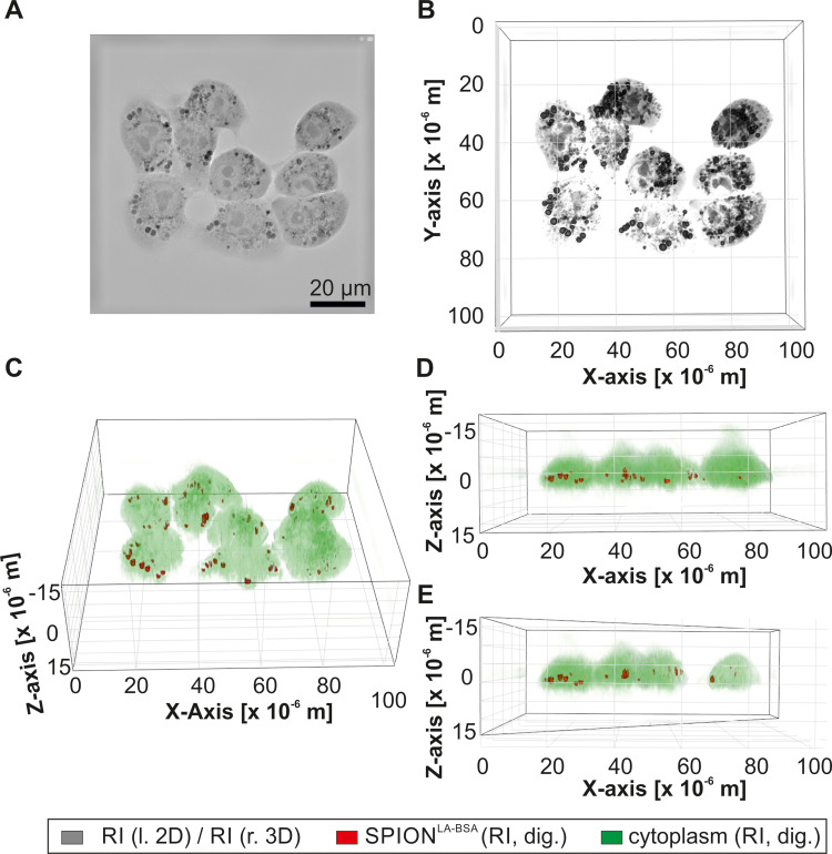
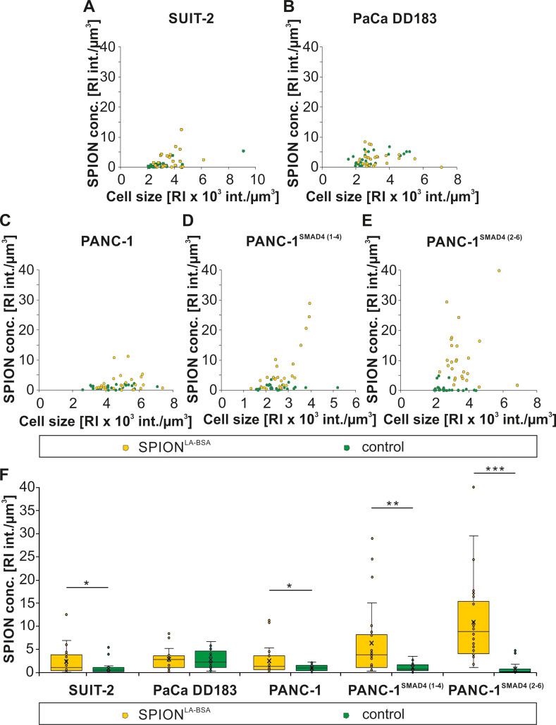
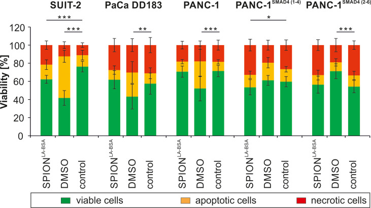
Results: Live cell imaging by 3D holotomographic microscopy demonstrated cell-specific differences in intracellular nanoparticle uptake in different pancreatic cell lines. Thus, treatment of PANC-1SMAD4 (1-4) and PANC-1SMAD4 (2-6) with SPIONs resulted in a significant increase in number of areas with higher RI, whereas in PANC-1, SUIT-2 and PaCa DD183, only a minimal increase of spots with high RI was observed. The increase in areas with high RI was in accordance with the SPION content determined by quantitative iron measurements using AES. In contrast, determination of the SPION amount by flow cytometry was strongly cell type-dependent and did not allow the discrimination between intracellular and membrane-bound SPIONs. However, flow cytometry is a very rapid and reliable method to assess the cellular toxicity and allows an estimation of the cell-associated SPION content.
Conclusion: Holotomographic 3D microscopy is a useful method to distinguish between intracellular and membrane-associated particles. Thus, it provides a valuable tool for scientists to evaluate the cellular localization and the particle load, which facilitates prediction of potential toxicity and efficiency of nanoparticles for medical applications.
期刊介绍:
Nanotechnology, Science and Applications is an international, peer-reviewed, Open Access journal that focuses on the science of nanotechnology in a wide range of industrial and academic applications. The journal is characterized by the rapid reporting of reviews, original research, and application studies across all sectors, including engineering, optics, bio-medicine, cosmetics, textiles, resource sustainability and science. Applied research into nano-materials, particles, nano-structures and fabrication, diagnostics and analytics, drug delivery and toxicology constitute the primary direction of the journal.


 分享
分享
 求助内容:
求助内容: 应助结果提醒方式:
 扫码关注我们
