Cezary Marcinkiewicz, Peter I Lelkes, Mark Sternberg, Giora Z Feuerstein
{"title":"荧光金刚石颗粒FDP-NV-800nm对人脐静脉原代细胞和人肝细胞系HepG-2体外基本生化功能的影响(六):急性生物相容性研究","authors":"Cezary Marcinkiewicz, Peter I Lelkes, Mark Sternberg, Giora Z Feuerstein","doi":"10.2147/NSA.S268107","DOIUrl":null,"url":null,"abstract":"<p><strong>Background: </strong>Recently, we reported the safety and biocompatibility of fluorescent diamond particles, FDP-NV-Z-800nm (FDP-NV) injected intravenously into rats, where no morbidity and mortality were noted over a period of 3 months. The acute effects of FDP-NV-800nm particles on cultured human endothelial and hepatic cells remain unexplored.</p><p><strong>Purpose: </strong>In this study, we aimed to explore select cellular and biochemical functions in cultured human umbilical endothelial cells (HUVEC) and a human hepatic cancer cell line (HepG-2) exposed to FDP-NV-800 in vitro at exposure levels within the pharmacokinetics (Cmax and the nadir) previously reported in vivo.</p><p><strong>Methods: </strong>Diverse cellular and biochemical functions were monitored, which cumulatively can provide insights into some vital cellular functions. Cell proliferation and migration were assessed by quantitative microscopy. Mitochondrial metabolic functions were tested by the MTT assay, and cytosolic esterase activity was studied by the calcein AM assay. Chaperons (CHOP), BiP and apoptosis (caspase-3 activation) were monitored by using Western blot (WB). MAPK Erk1/2 signaling was assessed by the detection of the phosphorylated form of the protein (P-Erk 1/2) and its translocation into the cell nucleus.</p><p><strong>Results: </strong>At all concentrations tested (0.001-0.1mg/mL), FDP-NV did not affect any of the biomarkers of cell integrity of HepG2 cells. In contrast, the proliferation of HUVEC was affected at the highest concentration tested (0.1mg/mL, C<sub>max</sub>). Exposure of HUVEC to (0.01 mg/mL) FDP-NV had a mild-moderate effect on cell proliferation as evident in the MTT assay and was absent when proliferation was assessed by direct cell counting or by using the calcein AM assays. In both cell types, exposure to the highest concentration (0.1 mg/mL) of FDP-NV did neither affect FBS-stimulated cell signaling (MAPK Erk1/2 phosphorylation) nor did it activate of Caspase 3.</p><p><strong>Conclusion: </strong>Our data suggest that FDP-NV-800nm are largely biocompatible with HepG-2 cells proliferation within the pharmacokinetic data reported previously. In contrast, HUVEC proliferation at the highest exposure dose (0.1 mg/mL) responded adversely with respect to several biomarkers of cell integrity. However, since the C<sub>max</sub> levels are very short-living, the risk for endothelial injury is likely minimal for slow rate cell proliferation such as endothelial cells.</p>","PeriodicalId":18881,"journal":{"name":"Nanotechnology, Science and Applications","volume":"13 ","pages":"103-118"},"PeriodicalIF":7.6000,"publicationDate":"2020-10-06","publicationTypes":"Journal Article","fieldsOfStudy":null,"isOpenAccess":false,"openAccessPdf":"https://sci-hub-pdf.com/10.2147/NSA.S268107","citationCount":"0","resultStr":"{\"title\":\"Effects of Fluorescent Diamond Particles FDP-NV-800nm on Essential Biochemical Functions of Primary Human Umbilical Vein Cells and Human Hepatic Cell Line, HepG-2 in vitro (Part VI): Acute Biocompatibility Studies.\",\"authors\":\"Cezary Marcinkiewicz, Peter I Lelkes, Mark Sternberg, Giora Z Feuerstein\",\"doi\":\"10.2147/NSA.S268107\",\"DOIUrl\":null,\"url\":null,\"abstract\":\"<p><strong>Background: </strong>Recently, we reported the safety and biocompatibility of fluorescent diamond particles, FDP-NV-Z-800nm (FDP-NV) injected intravenously into rats, where no morbidity and mortality were noted over a period of 3 months. The acute effects of FDP-NV-800nm particles on cultured human endothelial and hepatic cells remain unexplored.</p><p><strong>Purpose: </strong>In this study, we aimed to explore select cellular and biochemical functions in cultured human umbilical endothelial cells (HUVEC) and a human hepatic cancer cell line (HepG-2) exposed to FDP-NV-800 in vitro at exposure levels within the pharmacokinetics (Cmax and the nadir) previously reported in vivo.</p><p><strong>Methods: </strong>Diverse cellular and biochemical functions were monitored, which cumulatively can provide insights into some vital cellular functions. Cell proliferation and migration were assessed by quantitative microscopy. Mitochondrial metabolic functions were tested by the MTT assay, and cytosolic esterase activity was studied by the calcein AM assay. Chaperons (CHOP), BiP and apoptosis (caspase-3 activation) were monitored by using Western blot (WB). MAPK Erk1/2 signaling was assessed by the detection of the phosphorylated form of the protein (P-Erk 1/2) and its translocation into the cell nucleus.</p><p><strong>Results: </strong>At all concentrations tested (0.001-0.1mg/mL), FDP-NV did not affect any of the biomarkers of cell integrity of HepG2 cells. In contrast, the proliferation of HUVEC was affected at the highest concentration tested (0.1mg/mL, C<sub>max</sub>). Exposure of HUVEC to (0.01 mg/mL) FDP-NV had a mild-moderate effect on cell proliferation as evident in the MTT assay and was absent when proliferation was assessed by direct cell counting or by using the calcein AM assays. In both cell types, exposure to the highest concentration (0.1 mg/mL) of FDP-NV did neither affect FBS-stimulated cell signaling (MAPK Erk1/2 phosphorylation) nor did it activate of Caspase 3.</p><p><strong>Conclusion: </strong>Our data suggest that FDP-NV-800nm are largely biocompatible with HepG-2 cells proliferation within the pharmacokinetic data reported previously. In contrast, HUVEC proliferation at the highest exposure dose (0.1 mg/mL) responded adversely with respect to several biomarkers of cell integrity. However, since the C<sub>max</sub> levels are very short-living, the risk for endothelial injury is likely minimal for slow rate cell proliferation such as endothelial cells.</p>\",\"PeriodicalId\":18881,\"journal\":{\"name\":\"Nanotechnology, Science and Applications\",\"volume\":\"13 \",\"pages\":\"103-118\"},\"PeriodicalIF\":7.6000,\"publicationDate\":\"2020-10-06\",\"publicationTypes\":\"Journal Article\",\"fieldsOfStudy\":null,\"isOpenAccess\":false,\"openAccessPdf\":\"https://sci-hub-pdf.com/10.2147/NSA.S268107\",\"citationCount\":\"0\",\"resultStr\":null,\"platform\":\"Semanticscholar\",\"paperid\":null,\"PeriodicalName\":\"Nanotechnology, Science and Applications\",\"FirstCategoryId\":\"1085\",\"ListUrlMain\":\"https://doi.org/10.2147/NSA.S268107\",\"RegionNum\":0,\"RegionCategory\":null,\"ArticlePicture\":[],\"TitleCN\":null,\"AbstractTextCN\":null,\"PMCID\":null,\"EPubDate\":\"2020/1/1 0:00:00\",\"PubModel\":\"eCollection\",\"JCR\":\"Q2\",\"JCRName\":\"NANOSCIENCE & NANOTECHNOLOGY\",\"Score\":null,\"Total\":0}","platform":"Semanticscholar","paperid":null,"PeriodicalName":"Nanotechnology, Science and Applications","FirstCategoryId":"1085","ListUrlMain":"https://doi.org/10.2147/NSA.S268107","RegionNum":0,"RegionCategory":null,"ArticlePicture":[],"TitleCN":null,"AbstractTextCN":null,"PMCID":null,"EPubDate":"2020/1/1 0:00:00","PubModel":"eCollection","JCR":"Q2","JCRName":"NANOSCIENCE & NANOTECHNOLOGY","Score":null,"Total":0}
引用次数: 0
摘要
背景:最近,我们报道了FDP-NV- z -800nm荧光金刚石颗粒(FDP-NV)静脉注射大鼠的安全性和生物相容性,在3个月的时间里没有出现发病率和死亡率。FDP-NV-800nm颗粒对培养的人内皮细胞和肝细胞的急性作用尚未研究。目的:在本研究中,我们旨在探讨体外培养的人脐内皮细胞(HUVEC)和人肝癌细胞系(HepG-2)暴露于FDP-NV-800的细胞和生化功能,暴露水平在体内的药代动力学(Cmax和最低点)范围内。方法:对不同的细胞和生化功能进行监测,从而对一些重要的细胞功能有深入的了解。定量显微镜观察细胞增殖和迁移情况。MTT法测定线粒体代谢功能,钙黄蛋白AM法测定胞质酯酶活性。Western blot (WB)检测Chaperons (CHOP)、BiP和凋亡(caspase-3活化)。MAPK Erk1/2信号通过检测蛋白磷酸化形式(P-Erk 1/2)及其在细胞核中的易位来评估。结果:在所有浓度(0.001 ~ 0.1mg/mL)下,FDP-NV均未影响HepG2细胞完整性的任何生物标志物。而在最高浓度(0.1mg/mL, Cmax)时,HUVEC的增殖受到影响。在MTT试验中,HUVEC暴露于(0.01 mg/mL) FDP-NV对细胞增殖有轻度-中度影响,而通过直接细胞计数或使用钙黄蛋白AM试验评估增殖时则不存在这种影响。在两种细胞类型中,暴露于最高浓度(0.1 mg/mL)的FDP-NV既不影响fbs刺激的细胞信号传导(MAPK Erk1/2磷酸化),也不激活Caspase 3。结论:我们的数据表明,FDP-NV-800nm与HepG-2细胞增殖具有很大的生物相容性,符合先前报道的药代动力学数据。相反,在最高暴露剂量(0.1 mg/mL)下,HUVEC增殖对几种细胞完整性的生物标志物有不利反应。然而,由于Cmax水平的存在时间非常短,对于增殖缓慢的细胞(如内皮细胞),内皮损伤的风险可能很小。
Effects of Fluorescent Diamond Particles FDP-NV-800nm on Essential Biochemical Functions of Primary Human Umbilical Vein Cells and Human Hepatic Cell Line, HepG-2 in vitro (Part VI): Acute Biocompatibility Studies.
Background: Recently, we reported the safety and biocompatibility of fluorescent diamond particles, FDP-NV-Z-800nm (FDP-NV) injected intravenously into rats, where no morbidity and mortality were noted over a period of 3 months. The acute effects of FDP-NV-800nm particles on cultured human endothelial and hepatic cells remain unexplored.
Purpose: In this study, we aimed to explore select cellular and biochemical functions in cultured human umbilical endothelial cells (HUVEC) and a human hepatic cancer cell line (HepG-2) exposed to FDP-NV-800 in vitro at exposure levels within the pharmacokinetics (Cmax and the nadir) previously reported in vivo.
Methods: Diverse cellular and biochemical functions were monitored, which cumulatively can provide insights into some vital cellular functions. Cell proliferation and migration were assessed by quantitative microscopy. Mitochondrial metabolic functions were tested by the MTT assay, and cytosolic esterase activity was studied by the calcein AM assay. Chaperons (CHOP), BiP and apoptosis (caspase-3 activation) were monitored by using Western blot (WB). MAPK Erk1/2 signaling was assessed by the detection of the phosphorylated form of the protein (P-Erk 1/2) and its translocation into the cell nucleus.
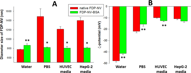
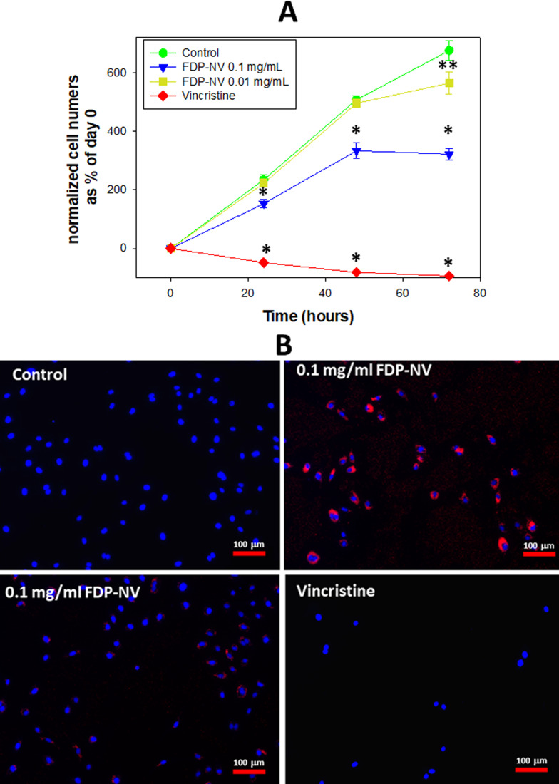
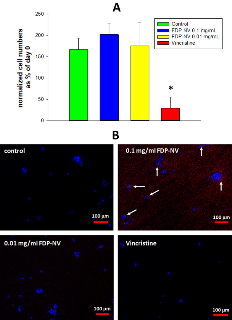
Results: At all concentrations tested (0.001-0.1mg/mL), FDP-NV did not affect any of the biomarkers of cell integrity of HepG2 cells. In contrast, the proliferation of HUVEC was affected at the highest concentration tested (0.1mg/mL, Cmax). Exposure of HUVEC to (0.01 mg/mL) FDP-NV had a mild-moderate effect on cell proliferation as evident in the MTT assay and was absent when proliferation was assessed by direct cell counting or by using the calcein AM assays. In both cell types, exposure to the highest concentration (0.1 mg/mL) of FDP-NV did neither affect FBS-stimulated cell signaling (MAPK Erk1/2 phosphorylation) nor did it activate of Caspase 3.
Conclusion: Our data suggest that FDP-NV-800nm are largely biocompatible with HepG-2 cells proliferation within the pharmacokinetic data reported previously. In contrast, HUVEC proliferation at the highest exposure dose (0.1 mg/mL) responded adversely with respect to several biomarkers of cell integrity. However, since the Cmax levels are very short-living, the risk for endothelial injury is likely minimal for slow rate cell proliferation such as endothelial cells.
期刊介绍:
Nanotechnology, Science and Applications is an international, peer-reviewed, Open Access journal that focuses on the science of nanotechnology in a wide range of industrial and academic applications. The journal is characterized by the rapid reporting of reviews, original research, and application studies across all sectors, including engineering, optics, bio-medicine, cosmetics, textiles, resource sustainability and science. Applied research into nano-materials, particles, nano-structures and fabrication, diagnostics and analytics, drug delivery and toxicology constitute the primary direction of the journal.


 分享
分享
 求助内容:
求助内容: 应助结果提醒方式:
 扫码关注我们
