{"title":"石蜡切片铁组织化学检测的简化方法:中枢神经系统组织细胞内铁沉积。","authors":"Steven M LeVine, Hao Zhu, Sarah E Tague","doi":"10.1177/1759091420982169","DOIUrl":null,"url":null,"abstract":"<p><p>Although all cells contain iron, most histochemical methods fail to reveal the presence of iron within many cells of the central nervous system (CNS), particularly neurons. Previously, a sensitive method was developed that limited the extraction of iron in paraffin sections, and this method revealed staining within neurons. However, the staining was often too robust making it difficult to discern discrete intracellular structures. In 1970, a study incorporated acetone in an iron histochemical procedure to facilitate the demarcation of staining features. In the present study, both acetone and limits to iron extraction were included in a simplified staining procedure. This procedure was applied to paraffin sections of CNS tissue from CISD2 deficient and littermate control mice. Discrete nuclear and cytoplasmic staining features were detected in all mice. Although widely present in neurons, punctate cytoplasmic staining was particularly prominent in large neurons within the hindbrain. Evaluation of extended depth of focus images, from serial focal planes, revealed numerous stained cytoplasmic structures. Additionally, the simplified staining procedure was applied to paraffin sections from Alzheimer's disease and control cases. Despite suboptimal processing conditions compared to mouse tissue, discrete staining of cytoplasmic structures was revealed in some neurons, although many other neurons had nondescript staining features. In addition, initial findings revealed iron deposited within some vessels from patients with Alzheimer's disease. In summary, since paraffin sections are commonly used for histological preparations, this simplified histochemical procedure could facilitate the study of iron in various CNS conditions by revealing staining details often missed by other procedures.</p>","PeriodicalId":8616,"journal":{"name":"ASN NEURO","volume":" ","pages":"1759091420982169"},"PeriodicalIF":3.7000,"publicationDate":"2021-01-01","publicationTypes":"Journal Article","fieldsOfStudy":null,"isOpenAccess":false,"openAccessPdf":"https://sci-hub-pdf.com/10.1177/1759091420982169","citationCount":"1","resultStr":"{\"title\":\"A Simplified Method for the Histochemical Detection of Iron in Paraffin Sections: Intracellular Iron Deposits in Central Nervous System Tissue.\",\"authors\":\"Steven M LeVine, Hao Zhu, Sarah E Tague\",\"doi\":\"10.1177/1759091420982169\",\"DOIUrl\":null,\"url\":null,\"abstract\":\"<p><p>Although all cells contain iron, most histochemical methods fail to reveal the presence of iron within many cells of the central nervous system (CNS), particularly neurons. Previously, a sensitive method was developed that limited the extraction of iron in paraffin sections, and this method revealed staining within neurons. However, the staining was often too robust making it difficult to discern discrete intracellular structures. In 1970, a study incorporated acetone in an iron histochemical procedure to facilitate the demarcation of staining features. In the present study, both acetone and limits to iron extraction were included in a simplified staining procedure. This procedure was applied to paraffin sections of CNS tissue from CISD2 deficient and littermate control mice. Discrete nuclear and cytoplasmic staining features were detected in all mice. Although widely present in neurons, punctate cytoplasmic staining was particularly prominent in large neurons within the hindbrain. Evaluation of extended depth of focus images, from serial focal planes, revealed numerous stained cytoplasmic structures. Additionally, the simplified staining procedure was applied to paraffin sections from Alzheimer's disease and control cases. Despite suboptimal processing conditions compared to mouse tissue, discrete staining of cytoplasmic structures was revealed in some neurons, although many other neurons had nondescript staining features. In addition, initial findings revealed iron deposited within some vessels from patients with Alzheimer's disease. In summary, since paraffin sections are commonly used for histological preparations, this simplified histochemical procedure could facilitate the study of iron in various CNS conditions by revealing staining details often missed by other procedures.</p>\",\"PeriodicalId\":8616,\"journal\":{\"name\":\"ASN NEURO\",\"volume\":\" \",\"pages\":\"1759091420982169\"},\"PeriodicalIF\":3.7000,\"publicationDate\":\"2021-01-01\",\"publicationTypes\":\"Journal Article\",\"fieldsOfStudy\":null,\"isOpenAccess\":false,\"openAccessPdf\":\"https://sci-hub-pdf.com/10.1177/1759091420982169\",\"citationCount\":\"1\",\"resultStr\":null,\"platform\":\"Semanticscholar\",\"paperid\":null,\"PeriodicalName\":\"ASN NEURO\",\"FirstCategoryId\":\"3\",\"ListUrlMain\":\"https://doi.org/10.1177/1759091420982169\",\"RegionNum\":4,\"RegionCategory\":\"医学\",\"ArticlePicture\":[],\"TitleCN\":null,\"AbstractTextCN\":null,\"PMCID\":null,\"EPubDate\":\"\",\"PubModel\":\"\",\"JCR\":\"Q2\",\"JCRName\":\"NEUROSCIENCES\",\"Score\":null,\"Total\":0}","platform":"Semanticscholar","paperid":null,"PeriodicalName":"ASN NEURO","FirstCategoryId":"3","ListUrlMain":"https://doi.org/10.1177/1759091420982169","RegionNum":4,"RegionCategory":"医学","ArticlePicture":[],"TitleCN":null,"AbstractTextCN":null,"PMCID":null,"EPubDate":"","PubModel":"","JCR":"Q2","JCRName":"NEUROSCIENCES","Score":null,"Total":0}
A Simplified Method for the Histochemical Detection of Iron in Paraffin Sections: Intracellular Iron Deposits in Central Nervous System Tissue.
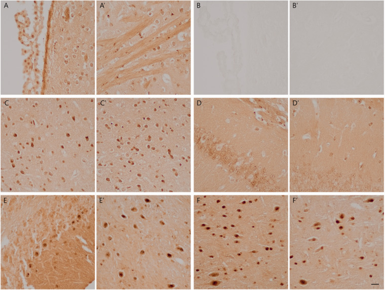
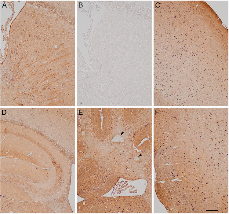
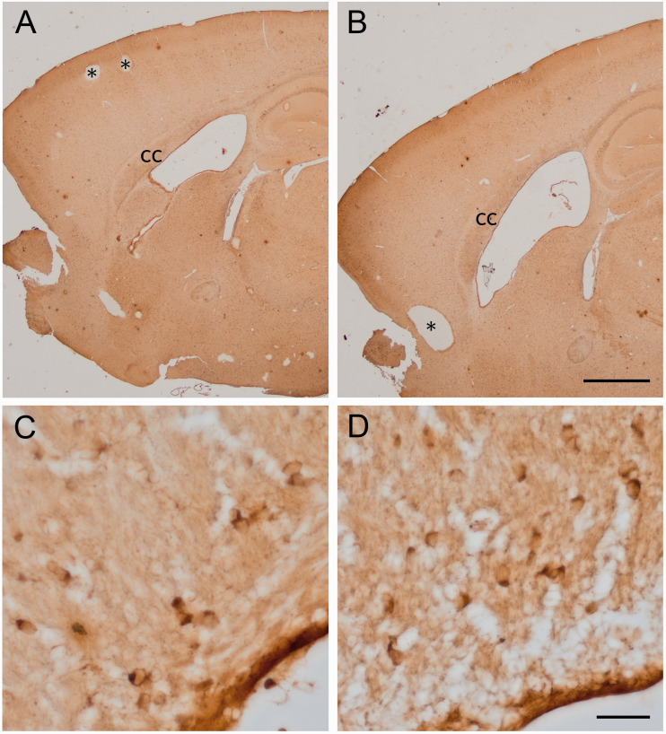
Although all cells contain iron, most histochemical methods fail to reveal the presence of iron within many cells of the central nervous system (CNS), particularly neurons. Previously, a sensitive method was developed that limited the extraction of iron in paraffin sections, and this method revealed staining within neurons. However, the staining was often too robust making it difficult to discern discrete intracellular structures. In 1970, a study incorporated acetone in an iron histochemical procedure to facilitate the demarcation of staining features. In the present study, both acetone and limits to iron extraction were included in a simplified staining procedure. This procedure was applied to paraffin sections of CNS tissue from CISD2 deficient and littermate control mice. Discrete nuclear and cytoplasmic staining features were detected in all mice. Although widely present in neurons, punctate cytoplasmic staining was particularly prominent in large neurons within the hindbrain. Evaluation of extended depth of focus images, from serial focal planes, revealed numerous stained cytoplasmic structures. Additionally, the simplified staining procedure was applied to paraffin sections from Alzheimer's disease and control cases. Despite suboptimal processing conditions compared to mouse tissue, discrete staining of cytoplasmic structures was revealed in some neurons, although many other neurons had nondescript staining features. In addition, initial findings revealed iron deposited within some vessels from patients with Alzheimer's disease. In summary, since paraffin sections are commonly used for histological preparations, this simplified histochemical procedure could facilitate the study of iron in various CNS conditions by revealing staining details often missed by other procedures.
期刊介绍:
ASN NEURO is an open access, peer-reviewed journal uniquely positioned to provide investigators with the most recent advances across the breadth of the cellular and molecular neurosciences. The official journal of the American Society for Neurochemistry, ASN NEURO is dedicated to the promotion, support, and facilitation of communication among cellular and molecular neuroscientists of all specializations.


 分享
分享
 求助内容:
求助内容: 应助结果提醒方式:
 扫码关注我们
