Corentin Malherbe, Bernard Crutzen, Jean Schrooyen, Giovanni Caruso, Frédéric Lecouvet, Christine Detrembleur, Thomas Schubert, Pierre-Louis Docquier
{"title":"骨肿瘤手术切除边缘的评估。","authors":"Corentin Malherbe, Bernard Crutzen, Jean Schrooyen, Giovanni Caruso, Frédéric Lecouvet, Christine Detrembleur, Thomas Schubert, Pierre-Louis Docquier","doi":"10.1155/2020/5289547","DOIUrl":null,"url":null,"abstract":"<p><p>Limb salvage surgery is now the preferred procedure for bone tumor surgery. To decrease the risk of local recurrence, it is crucial to obtain adequate resection margins. The obtained margins must be evaluated postoperatively because they influence what treatment is given subsequently when margins are not adequate (e.g., surgical revision and radiotherapy). The study aims to evaluate margin assessment of tumor specimen by MRI compared to conventional histology (to establish the viability of using MRI) and assess the accuracy of a patient-specific instrument when narrow margins were aimed. The resection margins in 12 consecutive patients that were operated on for bone tumor resection were prospectively analyzed using three methods: MRI of the resection specimen, macroscopic evaluation of specimen slices, and microscopic pathological evaluation. The assessments were qualitative (R0, R1, and R2) and quantitative (distance in mm). MRI, macroscopic, and microscopic margins generated similar results for both the qualitative (all resections were R0) and quantitative assessments. The median error in safe margins was 2 mm with a surgical guide (PSI) and 5 mm without a surgical guide. Local recurrences were not detected after a mean follow-up period of 3.7 years (range, 2.1-5 years); however, four patients died during the study. In conclusion, MRI is a valuable tool for assessing safe margins. When specimens are not available for pathological assessment (e.g., extracorporeally irradiated autograft or autoclaved autograft), MRI could be used to evaluate margins. In particular, when tumor volume is high, MRI could also help to focus the pathological examination on areas of concern.</p>","PeriodicalId":21431,"journal":{"name":"Sarcoma","volume":"2020 ","pages":"5289547"},"PeriodicalIF":0.0000,"publicationDate":"2020-12-10","publicationTypes":"Journal Article","fieldsOfStudy":null,"isOpenAccess":false,"openAccessPdf":"https://sci-hub-pdf.com/10.1155/2020/5289547","citationCount":"0","resultStr":"{\"title\":\"Assessment of Resection Margins in Bone Tumor Surgery.\",\"authors\":\"Corentin Malherbe, Bernard Crutzen, Jean Schrooyen, Giovanni Caruso, Frédéric Lecouvet, Christine Detrembleur, Thomas Schubert, Pierre-Louis Docquier\",\"doi\":\"10.1155/2020/5289547\",\"DOIUrl\":null,\"url\":null,\"abstract\":\"<p><p>Limb salvage surgery is now the preferred procedure for bone tumor surgery. To decrease the risk of local recurrence, it is crucial to obtain adequate resection margins. The obtained margins must be evaluated postoperatively because they influence what treatment is given subsequently when margins are not adequate (e.g., surgical revision and radiotherapy). The study aims to evaluate margin assessment of tumor specimen by MRI compared to conventional histology (to establish the viability of using MRI) and assess the accuracy of a patient-specific instrument when narrow margins were aimed. The resection margins in 12 consecutive patients that were operated on for bone tumor resection were prospectively analyzed using three methods: MRI of the resection specimen, macroscopic evaluation of specimen slices, and microscopic pathological evaluation. The assessments were qualitative (R0, R1, and R2) and quantitative (distance in mm). MRI, macroscopic, and microscopic margins generated similar results for both the qualitative (all resections were R0) and quantitative assessments. The median error in safe margins was 2 mm with a surgical guide (PSI) and 5 mm without a surgical guide. Local recurrences were not detected after a mean follow-up period of 3.7 years (range, 2.1-5 years); however, four patients died during the study. In conclusion, MRI is a valuable tool for assessing safe margins. When specimens are not available for pathological assessment (e.g., extracorporeally irradiated autograft or autoclaved autograft), MRI could be used to evaluate margins. In particular, when tumor volume is high, MRI could also help to focus the pathological examination on areas of concern.</p>\",\"PeriodicalId\":21431,\"journal\":{\"name\":\"Sarcoma\",\"volume\":\"2020 \",\"pages\":\"5289547\"},\"PeriodicalIF\":0.0000,\"publicationDate\":\"2020-12-10\",\"publicationTypes\":\"Journal Article\",\"fieldsOfStudy\":null,\"isOpenAccess\":false,\"openAccessPdf\":\"https://sci-hub-pdf.com/10.1155/2020/5289547\",\"citationCount\":\"0\",\"resultStr\":null,\"platform\":\"Semanticscholar\",\"paperid\":null,\"PeriodicalName\":\"Sarcoma\",\"FirstCategoryId\":\"1085\",\"ListUrlMain\":\"https://doi.org/10.1155/2020/5289547\",\"RegionNum\":0,\"RegionCategory\":null,\"ArticlePicture\":[],\"TitleCN\":null,\"AbstractTextCN\":null,\"PMCID\":null,\"EPubDate\":\"2020/1/1 0:00:00\",\"PubModel\":\"eCollection\",\"JCR\":\"Q2\",\"JCRName\":\"Medicine\",\"Score\":null,\"Total\":0}","platform":"Semanticscholar","paperid":null,"PeriodicalName":"Sarcoma","FirstCategoryId":"1085","ListUrlMain":"https://doi.org/10.1155/2020/5289547","RegionNum":0,"RegionCategory":null,"ArticlePicture":[],"TitleCN":null,"AbstractTextCN":null,"PMCID":null,"EPubDate":"2020/1/1 0:00:00","PubModel":"eCollection","JCR":"Q2","JCRName":"Medicine","Score":null,"Total":0}
Assessment of Resection Margins in Bone Tumor Surgery.
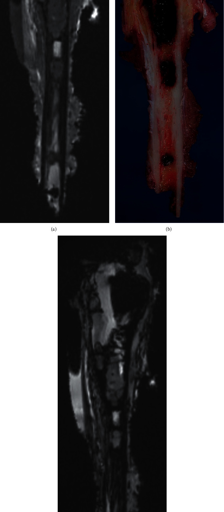
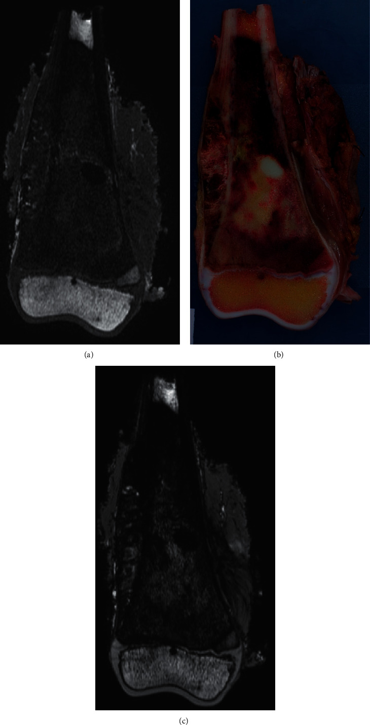
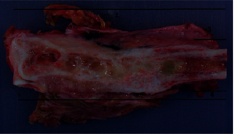
Limb salvage surgery is now the preferred procedure for bone tumor surgery. To decrease the risk of local recurrence, it is crucial to obtain adequate resection margins. The obtained margins must be evaluated postoperatively because they influence what treatment is given subsequently when margins are not adequate (e.g., surgical revision and radiotherapy). The study aims to evaluate margin assessment of tumor specimen by MRI compared to conventional histology (to establish the viability of using MRI) and assess the accuracy of a patient-specific instrument when narrow margins were aimed. The resection margins in 12 consecutive patients that were operated on for bone tumor resection were prospectively analyzed using three methods: MRI of the resection specimen, macroscopic evaluation of specimen slices, and microscopic pathological evaluation. The assessments were qualitative (R0, R1, and R2) and quantitative (distance in mm). MRI, macroscopic, and microscopic margins generated similar results for both the qualitative (all resections were R0) and quantitative assessments. The median error in safe margins was 2 mm with a surgical guide (PSI) and 5 mm without a surgical guide. Local recurrences were not detected after a mean follow-up period of 3.7 years (range, 2.1-5 years); however, four patients died during the study. In conclusion, MRI is a valuable tool for assessing safe margins. When specimens are not available for pathological assessment (e.g., extracorporeally irradiated autograft or autoclaved autograft), MRI could be used to evaluate margins. In particular, when tumor volume is high, MRI could also help to focus the pathological examination on areas of concern.
SarcomaMedicine-Radiology, Nuclear Medicine and Imaging
CiteScore
5.00
自引率
0.00%
发文量
15
审稿时长
14 weeks
期刊介绍:
Sarcoma is dedicated to publishing papers covering all aspects of connective tissue oncology research. It brings together work from scientists and clinicians carrying out a broad range of research in this field, including the basic sciences, molecular biology and pathology and the clinical sciences of epidemiology, surgery, radiotherapy and chemotherapy. High-quality papers concerning the entire range of bone and soft tissue sarcomas in both adults and children, including Kaposi"s sarcoma, are published as well as preclinical and animal studies. This journal provides a central forum for the description of advances in diagnosis, assessment and treatment of this rarely seen, but often mismanaged, group of patients.


 分享
分享
 求助内容:
求助内容: 应助结果提醒方式:
 扫码关注我们
