Yuchun Cai, Hongyue Zhan, Wenci Weng, Yao Wang, Pengxun Han, Xuewen Yu, Mumin Shao, Huili Sun
{"title":"氯胺醇胺通过抑制自噬改善糖尿病相关肌肉萎缩。","authors":"Yuchun Cai, Hongyue Zhan, Wenci Weng, Yao Wang, Pengxun Han, Xuewen Yu, Mumin Shao, Huili Sun","doi":"10.1186/s13395-021-00272-7","DOIUrl":null,"url":null,"abstract":"<p><strong>Background: </strong>Diabetes-related muscle wasting is one of the devastating complications of diabetes, which is associated with muscle autophagy due to insulin-mediated glucose starvation. However, treatment for diabetes-related muscle wasting is limited. Our previous study already found that niclosamide ethanolamine salt has the therapeutic effects on insulin deficiency of type 1 diabetes mice and muscle wasting induced by doxorubicin. Therefore, we aim to investigate the therapeutic effects of niclosamide ethanolamine salt on diabetes-induced muscle wasting and to explore whether the mechanism is associated with muscle autophagy.</p><p><strong>Methods: </strong>Type 1 diabetes mice were induced by intraperitoneal injection of streptozotocin, then were fed with regular diet supplemented with 10 g/kg niclosamide ethanolamine salt. The whole experiment lasted for 8 weeks. At the end of the study, grip strength, weights of tibialis anterior, gastrocnemius, soleus, and extensor digitorum longus muscle were measured. Tibialis anterior muscles stained with PAS were used for evaluating the fiber cross sectional area. Immunofluorescence analysis of myosin heavy chain expression in extensor digitorum longus and soleus muscle was used for determining the composition of the muscle fiber type. Electronic microscopy was applied to observe the autophagy in the atrophied muscle. Serum insulin levels and fasting blood glucose were also measured. Tissues of gastrocnemius muscle were used for detecting the expression of the proteins related to autophagy.</p><p><strong>Results: </strong>In this study, we found that niclosamide ethanolamine salt could ameliorate muscle atrophy in the type 1 diabetes mice as well, such as enhancing the declined grip strength, improving limb weight and increasing the numbers of glycolytic muscle fiber. Electron microscopy also confirmed that there did exist abundant autophagic vacuoles in the atrophied muscle of the type 1 diabetes mice. Specifically, niclosamide ethanolamine salt could reduce the over expression of autophagy-related proteins, including p-AMPK (Thr172), FoxO3a, p-ULK1 (Ser555), LC3B II, and p-p38 in gastrocnemius muscle of the type 1 diabetes mice.</p><p><strong>Conclusion: </strong>Niclosamide ethanolamine salt could ameliorate muscle wasting. The mechanisms underlying might be associated with inhibition of muscle autophagy.</p>","PeriodicalId":21747,"journal":{"name":"Skeletal Muscle","volume":" ","pages":"15"},"PeriodicalIF":4.4000,"publicationDate":"2021-06-09","publicationTypes":"Journal Article","fieldsOfStudy":null,"isOpenAccess":false,"openAccessPdf":"https://sci-hub-pdf.com/10.1186/s13395-021-00272-7","citationCount":"1","resultStr":"{\"title\":\"Niclosamide ethanolamine ameliorates diabetes-related muscle wasting by inhibiting autophagy.\",\"authors\":\"Yuchun Cai, Hongyue Zhan, Wenci Weng, Yao Wang, Pengxun Han, Xuewen Yu, Mumin Shao, Huili Sun\",\"doi\":\"10.1186/s13395-021-00272-7\",\"DOIUrl\":null,\"url\":null,\"abstract\":\"<p><strong>Background: </strong>Diabetes-related muscle wasting is one of the devastating complications of diabetes, which is associated with muscle autophagy due to insulin-mediated glucose starvation. However, treatment for diabetes-related muscle wasting is limited. Our previous study already found that niclosamide ethanolamine salt has the therapeutic effects on insulin deficiency of type 1 diabetes mice and muscle wasting induced by doxorubicin. Therefore, we aim to investigate the therapeutic effects of niclosamide ethanolamine salt on diabetes-induced muscle wasting and to explore whether the mechanism is associated with muscle autophagy.</p><p><strong>Methods: </strong>Type 1 diabetes mice were induced by intraperitoneal injection of streptozotocin, then were fed with regular diet supplemented with 10 g/kg niclosamide ethanolamine salt. The whole experiment lasted for 8 weeks. At the end of the study, grip strength, weights of tibialis anterior, gastrocnemius, soleus, and extensor digitorum longus muscle were measured. Tibialis anterior muscles stained with PAS were used for evaluating the fiber cross sectional area. Immunofluorescence analysis of myosin heavy chain expression in extensor digitorum longus and soleus muscle was used for determining the composition of the muscle fiber type. Electronic microscopy was applied to observe the autophagy in the atrophied muscle. Serum insulin levels and fasting blood glucose were also measured. Tissues of gastrocnemius muscle were used for detecting the expression of the proteins related to autophagy.</p><p><strong>Results: </strong>In this study, we found that niclosamide ethanolamine salt could ameliorate muscle atrophy in the type 1 diabetes mice as well, such as enhancing the declined grip strength, improving limb weight and increasing the numbers of glycolytic muscle fiber. Electron microscopy also confirmed that there did exist abundant autophagic vacuoles in the atrophied muscle of the type 1 diabetes mice. Specifically, niclosamide ethanolamine salt could reduce the over expression of autophagy-related proteins, including p-AMPK (Thr172), FoxO3a, p-ULK1 (Ser555), LC3B II, and p-p38 in gastrocnemius muscle of the type 1 diabetes mice.</p><p><strong>Conclusion: </strong>Niclosamide ethanolamine salt could ameliorate muscle wasting. The mechanisms underlying might be associated with inhibition of muscle autophagy.</p>\",\"PeriodicalId\":21747,\"journal\":{\"name\":\"Skeletal Muscle\",\"volume\":\" \",\"pages\":\"15\"},\"PeriodicalIF\":4.4000,\"publicationDate\":\"2021-06-09\",\"publicationTypes\":\"Journal Article\",\"fieldsOfStudy\":null,\"isOpenAccess\":false,\"openAccessPdf\":\"https://sci-hub-pdf.com/10.1186/s13395-021-00272-7\",\"citationCount\":\"1\",\"resultStr\":null,\"platform\":\"Semanticscholar\",\"paperid\":null,\"PeriodicalName\":\"Skeletal Muscle\",\"FirstCategoryId\":\"3\",\"ListUrlMain\":\"https://doi.org/10.1186/s13395-021-00272-7\",\"RegionNum\":2,\"RegionCategory\":\"医学\",\"ArticlePicture\":[],\"TitleCN\":null,\"AbstractTextCN\":null,\"PMCID\":null,\"EPubDate\":\"\",\"PubModel\":\"\",\"JCR\":\"Q2\",\"JCRName\":\"CELL BIOLOGY\",\"Score\":null,\"Total\":0}","platform":"Semanticscholar","paperid":null,"PeriodicalName":"Skeletal Muscle","FirstCategoryId":"3","ListUrlMain":"https://doi.org/10.1186/s13395-021-00272-7","RegionNum":2,"RegionCategory":"医学","ArticlePicture":[],"TitleCN":null,"AbstractTextCN":null,"PMCID":null,"EPubDate":"","PubModel":"","JCR":"Q2","JCRName":"CELL BIOLOGY","Score":null,"Total":0}
Niclosamide ethanolamine ameliorates diabetes-related muscle wasting by inhibiting autophagy.
Background: Diabetes-related muscle wasting is one of the devastating complications of diabetes, which is associated with muscle autophagy due to insulin-mediated glucose starvation. However, treatment for diabetes-related muscle wasting is limited. Our previous study already found that niclosamide ethanolamine salt has the therapeutic effects on insulin deficiency of type 1 diabetes mice and muscle wasting induced by doxorubicin. Therefore, we aim to investigate the therapeutic effects of niclosamide ethanolamine salt on diabetes-induced muscle wasting and to explore whether the mechanism is associated with muscle autophagy.
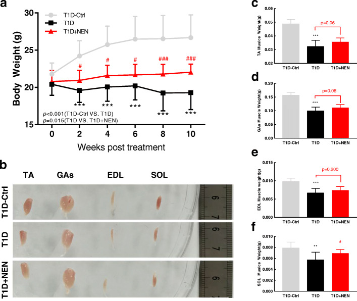
Methods: Type 1 diabetes mice were induced by intraperitoneal injection of streptozotocin, then were fed with regular diet supplemented with 10 g/kg niclosamide ethanolamine salt. The whole experiment lasted for 8 weeks. At the end of the study, grip strength, weights of tibialis anterior, gastrocnemius, soleus, and extensor digitorum longus muscle were measured. Tibialis anterior muscles stained with PAS were used for evaluating the fiber cross sectional area. Immunofluorescence analysis of myosin heavy chain expression in extensor digitorum longus and soleus muscle was used for determining the composition of the muscle fiber type. Electronic microscopy was applied to observe the autophagy in the atrophied muscle. Serum insulin levels and fasting blood glucose were also measured. Tissues of gastrocnemius muscle were used for detecting the expression of the proteins related to autophagy.
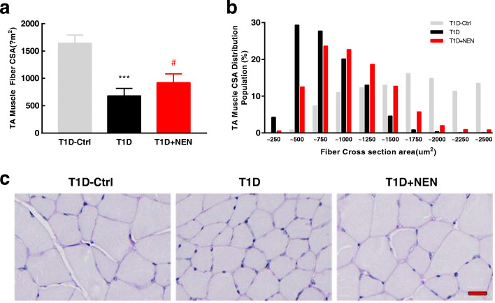
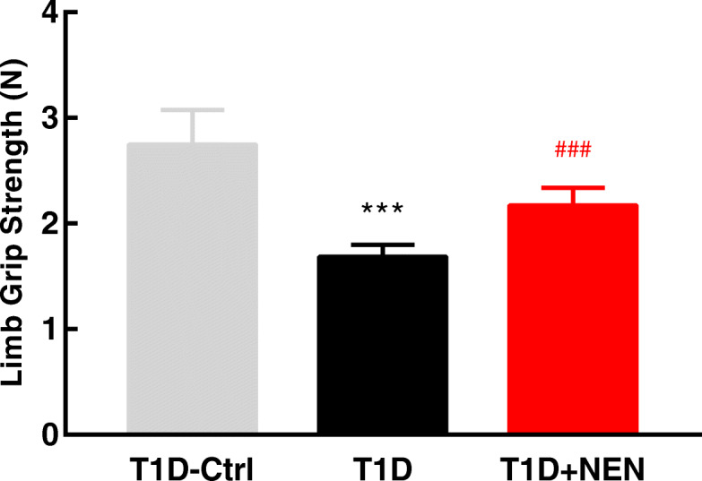
Results: In this study, we found that niclosamide ethanolamine salt could ameliorate muscle atrophy in the type 1 diabetes mice as well, such as enhancing the declined grip strength, improving limb weight and increasing the numbers of glycolytic muscle fiber. Electron microscopy also confirmed that there did exist abundant autophagic vacuoles in the atrophied muscle of the type 1 diabetes mice. Specifically, niclosamide ethanolamine salt could reduce the over expression of autophagy-related proteins, including p-AMPK (Thr172), FoxO3a, p-ULK1 (Ser555), LC3B II, and p-p38 in gastrocnemius muscle of the type 1 diabetes mice.
Conclusion: Niclosamide ethanolamine salt could ameliorate muscle wasting. The mechanisms underlying might be associated with inhibition of muscle autophagy.
期刊介绍:
The only open access journal in its field, Skeletal Muscle publishes novel, cutting-edge research and technological advancements that investigate the molecular mechanisms underlying the biology of skeletal muscle. Reflecting the breadth of research in this area, the journal welcomes manuscripts about the development, metabolism, the regulation of mass and function, aging, degeneration, dystrophy and regeneration of skeletal muscle, with an emphasis on understanding adult skeletal muscle, its maintenance, and its interactions with non-muscle cell types and regulatory modulators.
Main areas of interest include:
-differentiation of skeletal muscle-
atrophy and hypertrophy of skeletal muscle-
aging of skeletal muscle-
regeneration and degeneration of skeletal muscle-
biology of satellite and satellite-like cells-
dystrophic degeneration of skeletal muscle-
energy and glucose homeostasis in skeletal muscle-
non-dystrophic genetic diseases of skeletal muscle, such as Spinal Muscular Atrophy and myopathies-
maintenance of neuromuscular junctions-
roles of ryanodine receptors and calcium signaling in skeletal muscle-
roles of nuclear receptors in skeletal muscle-
roles of GPCRs and GPCR signaling in skeletal muscle-
other relevant aspects of skeletal muscle biology.
In addition, articles on translational clinical studies that address molecular and cellular mechanisms of skeletal muscle will be published. Case reports are also encouraged for submission.
Skeletal Muscle reflects the breadth of research on skeletal muscle and bridges gaps between diverse areas of science for example cardiac cell biology and neurobiology, which share common features with respect to cell differentiation, excitatory membranes, cell-cell communication, and maintenance. Suitable articles are model and mechanism-driven, and apply statistical principles where appropriate; purely descriptive studies are of lesser interest.


 分享
分享
 求助内容:
求助内容: 应助结果提醒方式:
 扫码关注我们
