Navjot Guru, Paula Demétrio De Souza França, Giacomo Pirovano, Cien Huang, Snehal G Patel, Thomas Reiner
{"title":"[18F]PARPi成像不受体外HPV状态的影响。","authors":"Navjot Guru, Paula Demétrio De Souza França, Giacomo Pirovano, Cien Huang, Snehal G Patel, Thomas Reiner","doi":"10.1155/2021/6641397","DOIUrl":null,"url":null,"abstract":"<p><strong>Background: </strong>Human papillomavirus- (HPV-) associated oropharyngeal squamous cell carcinomas (OPSCCs) are clinically and pathologically distinct from HPV-negative tumors. Here, we explore whether HPV affects functional biomarkers, including <i>γ</i>H2AX, RAD51, and PARP1. Moreover, the role of [<sup>18</sup>F]PARPi as a broadly applicable imaging tool for head and neck carcinomas is investigated.</p><p><strong>Methods: </strong>HPV-positive and HPV-negative cell lines were used to evaluate the <i>γ</i>H2AX, RAD51, and PARP1 expression with immunoblotting and immunofluorescence. Effects of external beam ionizing radiation were investigated <i>in vitro</i>, and survival was investigated via colony-formation assay. [<sup>18</sup>F]PARPi uptake experiments were performed on HPV-negative and HPV-positive cell lines to quantify PARP1 expression. PARP1 IHC and <i>γ</i>H2AX foci were quantified using patient-derived oropharyngeal tumor specimens.</p><p><strong>Results: </strong>Differences in DNA repair were detected, showing higher RAD51 and <i>γ</i>H2AX expression in HPV-positive cell lines. Clonogenic assays confirm HPV-positive cell lines to be significantly more radiosensitive. PARP1 expression levels were similar, irrespective of HPV status. Consequently, [<sup>18</sup>F]PARPi uptake assays demonstrated that this tracer is internalized in cell lines independently from their HPV status.</p><p><strong>Conclusion: </strong>The HPV status, often used clinically to stratify patients, did not affect PARP1 levels, suggesting that PARP imaging can be performed in both HPV-positive and HPV-negative patients. This study confirms that the PET imaging agent [<sup>18</sup>F]PARPi could serve as a general clinical tool for oropharyngeal cancer patients.</p>","PeriodicalId":2,"journal":{"name":"ACS Applied Bio Materials","volume":" ","pages":"6641397"},"PeriodicalIF":4.7000,"publicationDate":"2021-01-20","publicationTypes":"Journal Article","fieldsOfStudy":null,"isOpenAccess":false,"openAccessPdf":"https://www.ncbi.nlm.nih.gov/pmc/articles/PMC8205605/pdf/","citationCount":"0","resultStr":"{\"title\":\"[<sup>18</sup>F]PARPi Imaging Is Not Affected by HPV Status In Vitro.\",\"authors\":\"Navjot Guru, Paula Demétrio De Souza França, Giacomo Pirovano, Cien Huang, Snehal G Patel, Thomas Reiner\",\"doi\":\"10.1155/2021/6641397\",\"DOIUrl\":null,\"url\":null,\"abstract\":\"<p><strong>Background: </strong>Human papillomavirus- (HPV-) associated oropharyngeal squamous cell carcinomas (OPSCCs) are clinically and pathologically distinct from HPV-negative tumors. Here, we explore whether HPV affects functional biomarkers, including <i>γ</i>H2AX, RAD51, and PARP1. Moreover, the role of [<sup>18</sup>F]PARPi as a broadly applicable imaging tool for head and neck carcinomas is investigated.</p><p><strong>Methods: </strong>HPV-positive and HPV-negative cell lines were used to evaluate the <i>γ</i>H2AX, RAD51, and PARP1 expression with immunoblotting and immunofluorescence. Effects of external beam ionizing radiation were investigated <i>in vitro</i>, and survival was investigated via colony-formation assay. [<sup>18</sup>F]PARPi uptake experiments were performed on HPV-negative and HPV-positive cell lines to quantify PARP1 expression. PARP1 IHC and <i>γ</i>H2AX foci were quantified using patient-derived oropharyngeal tumor specimens.</p><p><strong>Results: </strong>Differences in DNA repair were detected, showing higher RAD51 and <i>γ</i>H2AX expression in HPV-positive cell lines. Clonogenic assays confirm HPV-positive cell lines to be significantly more radiosensitive. PARP1 expression levels were similar, irrespective of HPV status. Consequently, [<sup>18</sup>F]PARPi uptake assays demonstrated that this tracer is internalized in cell lines independently from their HPV status.</p><p><strong>Conclusion: </strong>The HPV status, often used clinically to stratify patients, did not affect PARP1 levels, suggesting that PARP imaging can be performed in both HPV-positive and HPV-negative patients. This study confirms that the PET imaging agent [<sup>18</sup>F]PARPi could serve as a general clinical tool for oropharyngeal cancer patients.</p>\",\"PeriodicalId\":2,\"journal\":{\"name\":\"ACS Applied Bio Materials\",\"volume\":\" \",\"pages\":\"6641397\"},\"PeriodicalIF\":4.7000,\"publicationDate\":\"2021-01-20\",\"publicationTypes\":\"Journal Article\",\"fieldsOfStudy\":null,\"isOpenAccess\":false,\"openAccessPdf\":\"https://www.ncbi.nlm.nih.gov/pmc/articles/PMC8205605/pdf/\",\"citationCount\":\"0\",\"resultStr\":null,\"platform\":\"Semanticscholar\",\"paperid\":null,\"PeriodicalName\":\"ACS Applied Bio Materials\",\"FirstCategoryId\":\"3\",\"ListUrlMain\":\"https://doi.org/10.1155/2021/6641397\",\"RegionNum\":0,\"RegionCategory\":null,\"ArticlePicture\":[],\"TitleCN\":null,\"AbstractTextCN\":null,\"PMCID\":null,\"EPubDate\":\"2021/1/1 0:00:00\",\"PubModel\":\"eCollection\",\"JCR\":\"Q2\",\"JCRName\":\"MATERIALS SCIENCE, BIOMATERIALS\",\"Score\":null,\"Total\":0}","platform":"Semanticscholar","paperid":null,"PeriodicalName":"ACS Applied Bio Materials","FirstCategoryId":"3","ListUrlMain":"https://doi.org/10.1155/2021/6641397","RegionNum":0,"RegionCategory":null,"ArticlePicture":[],"TitleCN":null,"AbstractTextCN":null,"PMCID":null,"EPubDate":"2021/1/1 0:00:00","PubModel":"eCollection","JCR":"Q2","JCRName":"MATERIALS SCIENCE, BIOMATERIALS","Score":null,"Total":0}
[18F]PARPi Imaging Is Not Affected by HPV Status In Vitro.
Background: Human papillomavirus- (HPV-) associated oropharyngeal squamous cell carcinomas (OPSCCs) are clinically and pathologically distinct from HPV-negative tumors. Here, we explore whether HPV affects functional biomarkers, including γH2AX, RAD51, and PARP1. Moreover, the role of [18F]PARPi as a broadly applicable imaging tool for head and neck carcinomas is investigated.
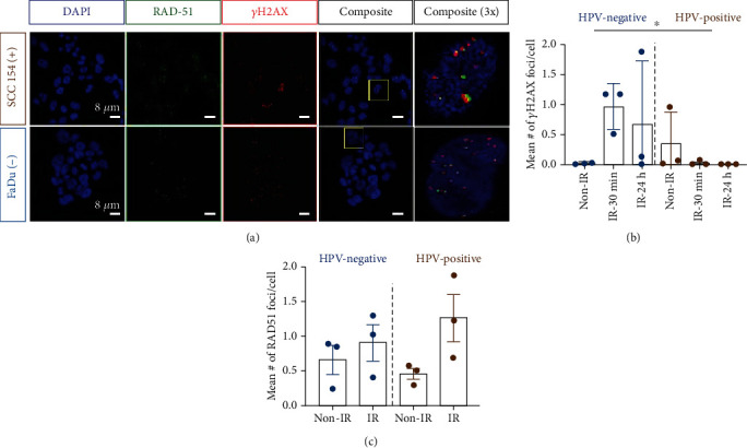
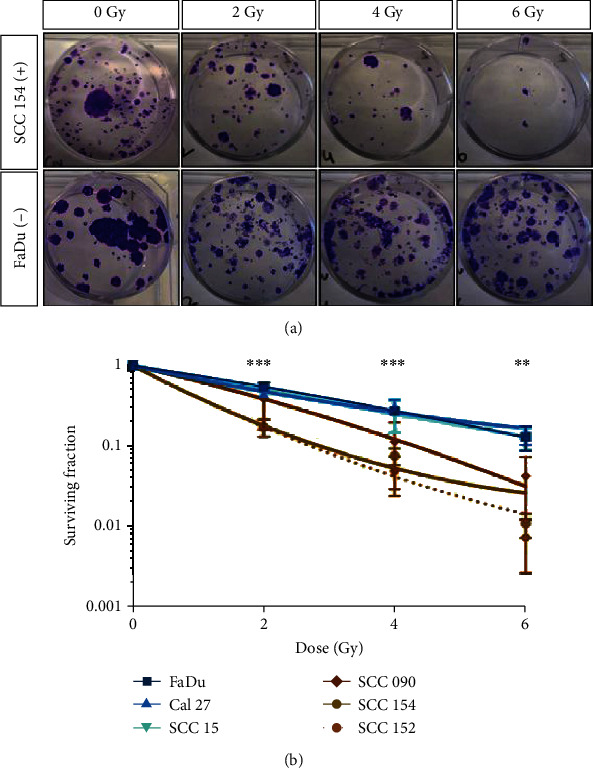
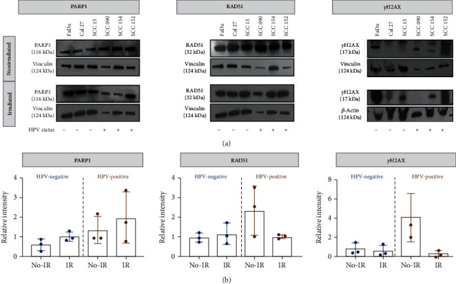
Methods: HPV-positive and HPV-negative cell lines were used to evaluate the γH2AX, RAD51, and PARP1 expression with immunoblotting and immunofluorescence. Effects of external beam ionizing radiation were investigated in vitro, and survival was investigated via colony-formation assay. [18F]PARPi uptake experiments were performed on HPV-negative and HPV-positive cell lines to quantify PARP1 expression. PARP1 IHC and γH2AX foci were quantified using patient-derived oropharyngeal tumor specimens.
Results: Differences in DNA repair were detected, showing higher RAD51 and γH2AX expression in HPV-positive cell lines. Clonogenic assays confirm HPV-positive cell lines to be significantly more radiosensitive. PARP1 expression levels were similar, irrespective of HPV status. Consequently, [18F]PARPi uptake assays demonstrated that this tracer is internalized in cell lines independently from their HPV status.
Conclusion: The HPV status, often used clinically to stratify patients, did not affect PARP1 levels, suggesting that PARP imaging can be performed in both HPV-positive and HPV-negative patients. This study confirms that the PET imaging agent [18F]PARPi could serve as a general clinical tool for oropharyngeal cancer patients.
期刊介绍:
ACS Applied Bio Materials is an interdisciplinary journal publishing original research covering all aspects of biomaterials and biointerfaces including and beyond the traditional biosensing, biomedical and therapeutic applications.
The journal is devoted to reports of new and original experimental and theoretical research of an applied nature that integrates knowledge in the areas of materials, engineering, physics, bioscience, and chemistry into important bio applications. The journal is specifically interested in work that addresses the relationship between structure and function and assesses the stability and degradation of materials under relevant environmental and biological conditions.


 分享
分享
 求助内容:
求助内容: 应助结果提醒方式:
 扫码关注我们
