Simone Krebs, Julia G Barasch, Robert J Young, Christian Grommes, Heiko Schöder
{"title":"原发性中枢神经系统淋巴瘤的正电子发射断层成像和磁共振成像--综述。","authors":"Simone Krebs, Julia G Barasch, Robert J Young, Christian Grommes, Heiko Schöder","doi":"10.21037/aol-20-52","DOIUrl":null,"url":null,"abstract":"<p><p>This review addresses the challenges of primary central nervous system (CNS) lymphoma diagnosis, assessment of treatment response, and detection of recurrence. Primary CNS lymphoma is a rare form of extra-nodal non-Hodgkin lymphoma that can involve brain, spinal cord, leptomeninges, and eyes. Primary CNS lymphoma lesions are most commonly confined to the white matter or deep cerebral structures such as basal ganglia and deep periventricular regions. Contrast-enhanced magnetic resonance imaging (MRI) is the standard diagnostic modality employed by neuro-oncologists. MRI often shows common morphological features such as a single or multiple uniformly well-enhancing lesions without necrosis but with moderate surrounding edema. Other brain tumors or inflammatory processes can show similar radiological patterns, making differential diagnosis difficult. [18F]-fluorodeoxyglucose (FDG) positron emission tomography (PET) has selected utility in cerebral lymphoma, especially in diagnosis. Primary CNS lymphoma can sometimes present with atypical findings on MRI and FDG PET, such as disseminated disease, non-enhancing or ring-like enhancing lesions. The complementary strengths of PET and MRI have led to the development of combined PET-MR systems, which in some cases may improve lesion characterization and detection. By highlighting active developments in this field, including advanced MRI sequences, novel radiotracers, and potential imaging biomarkers, we aim to spur interest in sophisticated imaging approaches.</p>","PeriodicalId":72224,"journal":{"name":"Annals of lymphoma","volume":"5 ","pages":""},"PeriodicalIF":0.0000,"publicationDate":"2021-06-01","publicationTypes":"Journal Article","fieldsOfStudy":null,"isOpenAccess":false,"openAccessPdf":"https://ftp.ncbi.nlm.nih.gov/pub/pmc/oa_pdf/da/55/nihms-1715614.PMC8248935.pdf","citationCount":"0","resultStr":"{\"title\":\"Positron emission tomography and magnetic resonance imaging in primary central nervous system lymphoma-a narrative review.\",\"authors\":\"Simone Krebs, Julia G Barasch, Robert J Young, Christian Grommes, Heiko Schöder\",\"doi\":\"10.21037/aol-20-52\",\"DOIUrl\":null,\"url\":null,\"abstract\":\"<p><p>This review addresses the challenges of primary central nervous system (CNS) lymphoma diagnosis, assessment of treatment response, and detection of recurrence. Primary CNS lymphoma is a rare form of extra-nodal non-Hodgkin lymphoma that can involve brain, spinal cord, leptomeninges, and eyes. Primary CNS lymphoma lesions are most commonly confined to the white matter or deep cerebral structures such as basal ganglia and deep periventricular regions. Contrast-enhanced magnetic resonance imaging (MRI) is the standard diagnostic modality employed by neuro-oncologists. MRI often shows common morphological features such as a single or multiple uniformly well-enhancing lesions without necrosis but with moderate surrounding edema. Other brain tumors or inflammatory processes can show similar radiological patterns, making differential diagnosis difficult. [18F]-fluorodeoxyglucose (FDG) positron emission tomography (PET) has selected utility in cerebral lymphoma, especially in diagnosis. Primary CNS lymphoma can sometimes present with atypical findings on MRI and FDG PET, such as disseminated disease, non-enhancing or ring-like enhancing lesions. The complementary strengths of PET and MRI have led to the development of combined PET-MR systems, which in some cases may improve lesion characterization and detection. By highlighting active developments in this field, including advanced MRI sequences, novel radiotracers, and potential imaging biomarkers, we aim to spur interest in sophisticated imaging approaches.</p>\",\"PeriodicalId\":72224,\"journal\":{\"name\":\"Annals of lymphoma\",\"volume\":\"5 \",\"pages\":\"\"},\"PeriodicalIF\":0.0000,\"publicationDate\":\"2021-06-01\",\"publicationTypes\":\"Journal Article\",\"fieldsOfStudy\":null,\"isOpenAccess\":false,\"openAccessPdf\":\"https://ftp.ncbi.nlm.nih.gov/pub/pmc/oa_pdf/da/55/nihms-1715614.PMC8248935.pdf\",\"citationCount\":\"0\",\"resultStr\":null,\"platform\":\"Semanticscholar\",\"paperid\":null,\"PeriodicalName\":\"Annals of lymphoma\",\"FirstCategoryId\":\"1085\",\"ListUrlMain\":\"https://doi.org/10.21037/aol-20-52\",\"RegionNum\":0,\"RegionCategory\":null,\"ArticlePicture\":[],\"TitleCN\":null,\"AbstractTextCN\":null,\"PMCID\":null,\"EPubDate\":\"2021/6/30 0:00:00\",\"PubModel\":\"Epub\",\"JCR\":\"\",\"JCRName\":\"\",\"Score\":null,\"Total\":0}","platform":"Semanticscholar","paperid":null,"PeriodicalName":"Annals of lymphoma","FirstCategoryId":"1085","ListUrlMain":"https://doi.org/10.21037/aol-20-52","RegionNum":0,"RegionCategory":null,"ArticlePicture":[],"TitleCN":null,"AbstractTextCN":null,"PMCID":null,"EPubDate":"2021/6/30 0:00:00","PubModel":"Epub","JCR":"","JCRName":"","Score":null,"Total":0}
Positron emission tomography and magnetic resonance imaging in primary central nervous system lymphoma-a narrative review.
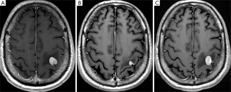
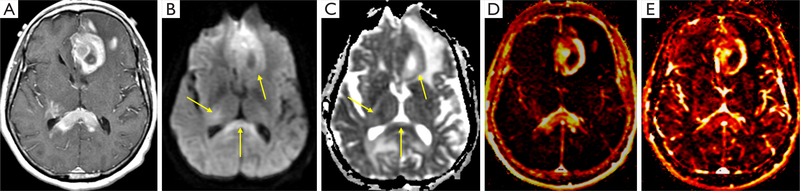
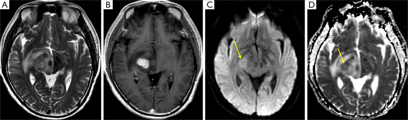
This review addresses the challenges of primary central nervous system (CNS) lymphoma diagnosis, assessment of treatment response, and detection of recurrence. Primary CNS lymphoma is a rare form of extra-nodal non-Hodgkin lymphoma that can involve brain, spinal cord, leptomeninges, and eyes. Primary CNS lymphoma lesions are most commonly confined to the white matter or deep cerebral structures such as basal ganglia and deep periventricular regions. Contrast-enhanced magnetic resonance imaging (MRI) is the standard diagnostic modality employed by neuro-oncologists. MRI often shows common morphological features such as a single or multiple uniformly well-enhancing lesions without necrosis but with moderate surrounding edema. Other brain tumors or inflammatory processes can show similar radiological patterns, making differential diagnosis difficult. [18F]-fluorodeoxyglucose (FDG) positron emission tomography (PET) has selected utility in cerebral lymphoma, especially in diagnosis. Primary CNS lymphoma can sometimes present with atypical findings on MRI and FDG PET, such as disseminated disease, non-enhancing or ring-like enhancing lesions. The complementary strengths of PET and MRI have led to the development of combined PET-MR systems, which in some cases may improve lesion characterization and detection. By highlighting active developments in this field, including advanced MRI sequences, novel radiotracers, and potential imaging biomarkers, we aim to spur interest in sophisticated imaging approaches.


 分享
分享

 求助内容:
求助内容: 应助结果提醒方式:
 扫码关注我们
