{"title":"全膝关节置换术中假体位置三维与二维评价的比较。","authors":"Osamu Tanifuji, Tomoharu Mochizuki, Hiroshi Yamagiwa, Takashi Sato, Satoshi Watanabe, Hiroki Hijikata, Hiroyuki Kawashima","doi":"10.1186/s43019-021-00106-2","DOIUrl":null,"url":null,"abstract":"<p><strong>Purpose: </strong>The purpose of this study was to evaluate the post-operative three-dimensional (3D) femoral and tibial component positions in total knee arthroplasty (TKA) by the same co-ordinates' system as for pre-operative planning and to compare it with a two-dimensional (2D) evaluation.</p><p><strong>Materials and methods: </strong>Sixty-five primary TKAs due to osteoarthritis were included. A computed tomography (CT) scan of the femur and tibia was obtained and pre-operative 3D planning was performed. Then, 3D and 2D post-operative evaluations of the component positions were performed. KneeCAS (LEXI, Inc., Tokyo, Japan), a lower-extremity alignment assessment system, was used for the 3D post-operative evaluation. Standard short-knee radiographs were used for the 2D post-operative evaluation. Differences between the pre-operative planning and post-operative coronal and sagittal alignment of components were investigated and compared with the results of the 3D and 2D evaluations.</p><p><strong>Results: </strong>According to the 3D evaluation, the difference between the pre-operative planning and actual post-operative sagittal alignment of the femoral component and the coronal and sagittal alignments of the tibial component were 2.6° ± 1.8°, 2.2° ± 1.8° and 3.2° ± 2.4°, respectively. Using the 2D evaluation, they were 1.9° ± 1.5°, 1.3° ± 1.2° and 1.8° ± 1.4°, making the difference in 3D evaluation significantly higher (p = 0.013, = 0.003 and < 0.001). For the sagittal alignment of the femoral component and the coronal and sagittal alignment of the tibial component, the outlier (> ± 3°) ratio for the 3D evaluation was also significantly higher than that of the 2D evaluation (p < 0.001, = 0.009 and < 0.001).</p><p><strong>Conclusions: </strong>The difference between the pre-operative planning and post-operative component alignment in the 3D evaluation is significantly higher than that of the 2D, even if the same cases have been evaluated. Two-dimensional evaluation may mask or underestimate the post-operative implant malposition. Three-dimensional evaluation using the same co-ordinates' system as for pre-operative planning is necessary to accurately evaluate the post-operative component position.</p>","PeriodicalId":17886,"journal":{"name":"Knee Surgery & Related Research","volume":"33 1","pages":"21"},"PeriodicalIF":4.4000,"publicationDate":"2021-07-13","publicationTypes":"Journal Article","fieldsOfStudy":null,"isOpenAccess":false,"openAccessPdf":"https://sci-hub-pdf.com/10.1186/s43019-021-00106-2","citationCount":"6","resultStr":"{\"title\":\"Comparison of post-operative three-dimensional and two-dimensional evaluation of component position for total knee arthroplasty.\",\"authors\":\"Osamu Tanifuji, Tomoharu Mochizuki, Hiroshi Yamagiwa, Takashi Sato, Satoshi Watanabe, Hiroki Hijikata, Hiroyuki Kawashima\",\"doi\":\"10.1186/s43019-021-00106-2\",\"DOIUrl\":null,\"url\":null,\"abstract\":\"<p><strong>Purpose: </strong>The purpose of this study was to evaluate the post-operative three-dimensional (3D) femoral and tibial component positions in total knee arthroplasty (TKA) by the same co-ordinates' system as for pre-operative planning and to compare it with a two-dimensional (2D) evaluation.</p><p><strong>Materials and methods: </strong>Sixty-five primary TKAs due to osteoarthritis were included. A computed tomography (CT) scan of the femur and tibia was obtained and pre-operative 3D planning was performed. Then, 3D and 2D post-operative evaluations of the component positions were performed. KneeCAS (LEXI, Inc., Tokyo, Japan), a lower-extremity alignment assessment system, was used for the 3D post-operative evaluation. Standard short-knee radiographs were used for the 2D post-operative evaluation. Differences between the pre-operative planning and post-operative coronal and sagittal alignment of components were investigated and compared with the results of the 3D and 2D evaluations.</p><p><strong>Results: </strong>According to the 3D evaluation, the difference between the pre-operative planning and actual post-operative sagittal alignment of the femoral component and the coronal and sagittal alignments of the tibial component were 2.6° ± 1.8°, 2.2° ± 1.8° and 3.2° ± 2.4°, respectively. Using the 2D evaluation, they were 1.9° ± 1.5°, 1.3° ± 1.2° and 1.8° ± 1.4°, making the difference in 3D evaluation significantly higher (p = 0.013, = 0.003 and < 0.001). For the sagittal alignment of the femoral component and the coronal and sagittal alignment of the tibial component, the outlier (> ± 3°) ratio for the 3D evaluation was also significantly higher than that of the 2D evaluation (p < 0.001, = 0.009 and < 0.001).</p><p><strong>Conclusions: </strong>The difference between the pre-operative planning and post-operative component alignment in the 3D evaluation is significantly higher than that of the 2D, even if the same cases have been evaluated. Two-dimensional evaluation may mask or underestimate the post-operative implant malposition. Three-dimensional evaluation using the same co-ordinates' system as for pre-operative planning is necessary to accurately evaluate the post-operative component position.</p>\",\"PeriodicalId\":17886,\"journal\":{\"name\":\"Knee Surgery & Related Research\",\"volume\":\"33 1\",\"pages\":\"21\"},\"PeriodicalIF\":4.4000,\"publicationDate\":\"2021-07-13\",\"publicationTypes\":\"Journal Article\",\"fieldsOfStudy\":null,\"isOpenAccess\":false,\"openAccessPdf\":\"https://sci-hub-pdf.com/10.1186/s43019-021-00106-2\",\"citationCount\":\"6\",\"resultStr\":null,\"platform\":\"Semanticscholar\",\"paperid\":null,\"PeriodicalName\":\"Knee Surgery & Related Research\",\"FirstCategoryId\":\"1085\",\"ListUrlMain\":\"https://doi.org/10.1186/s43019-021-00106-2\",\"RegionNum\":0,\"RegionCategory\":null,\"ArticlePicture\":[],\"TitleCN\":null,\"AbstractTextCN\":null,\"PMCID\":null,\"EPubDate\":\"\",\"PubModel\":\"\",\"JCR\":\"Q1\",\"JCRName\":\"ORTHOPEDICS\",\"Score\":null,\"Total\":0}","platform":"Semanticscholar","paperid":null,"PeriodicalName":"Knee Surgery & Related Research","FirstCategoryId":"1085","ListUrlMain":"https://doi.org/10.1186/s43019-021-00106-2","RegionNum":0,"RegionCategory":null,"ArticlePicture":[],"TitleCN":null,"AbstractTextCN":null,"PMCID":null,"EPubDate":"","PubModel":"","JCR":"Q1","JCRName":"ORTHOPEDICS","Score":null,"Total":0}
引用次数: 6
摘要
目的:本研究的目的是通过与术前计划相同的坐标系统评估全膝关节置换术(TKA)中股骨和胫骨部件的术后三维(3D)位置,并将其与二维(2D)评估进行比较。材料与方法:纳入65例原发性骨关节炎tka。获得股骨和胫骨的计算机断层扫描(CT),并进行术前3D计划。然后,进行三维和二维的术后部件位置评估。膝关节定位评估系统(LEXI, Inc., Tokyo, Japan)用于术后3D评估。使用标准短膝x线片进行术后2D评估。研究术前规划与术后椎体冠状和矢状对齐的差异,并与3D和2D评估结果进行比较。结果:根据三维评估,术前规划与实际术后股骨假体矢状位对齐与胫骨假体冠状位、矢状位对齐的差值分别为2.6°±1.8°、2.2°±1.8°和3.2°±2.4°。使用2D评估时,它们分别为1.9°±1.5°、1.3°±1.2°和1.8°±1.4°,使得3D评估的差异显著高于2D评估(p = 0.013、= 0.003和±3°),且3D评估的差异也显著高于2D评估(p结论:即使对相同病例进行评估,3D评估中术前计划和术后部件对齐的差异也显著高于2D评估。二维评估可能掩盖或低估术后种植体错位。使用与术前规划相同的坐标系进行三维评估是准确评估术后部件位置的必要条件。
Comparison of post-operative three-dimensional and two-dimensional evaluation of component position for total knee arthroplasty.
Purpose: The purpose of this study was to evaluate the post-operative three-dimensional (3D) femoral and tibial component positions in total knee arthroplasty (TKA) by the same co-ordinates' system as for pre-operative planning and to compare it with a two-dimensional (2D) evaluation.
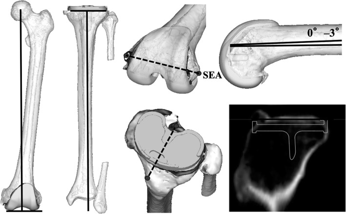
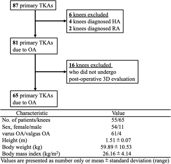
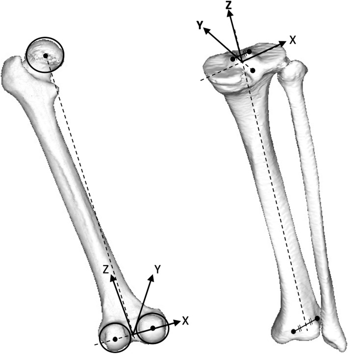
Materials and methods: Sixty-five primary TKAs due to osteoarthritis were included. A computed tomography (CT) scan of the femur and tibia was obtained and pre-operative 3D planning was performed. Then, 3D and 2D post-operative evaluations of the component positions were performed. KneeCAS (LEXI, Inc., Tokyo, Japan), a lower-extremity alignment assessment system, was used for the 3D post-operative evaluation. Standard short-knee radiographs were used for the 2D post-operative evaluation. Differences between the pre-operative planning and post-operative coronal and sagittal alignment of components were investigated and compared with the results of the 3D and 2D evaluations.
Results: According to the 3D evaluation, the difference between the pre-operative planning and actual post-operative sagittal alignment of the femoral component and the coronal and sagittal alignments of the tibial component were 2.6° ± 1.8°, 2.2° ± 1.8° and 3.2° ± 2.4°, respectively. Using the 2D evaluation, they were 1.9° ± 1.5°, 1.3° ± 1.2° and 1.8° ± 1.4°, making the difference in 3D evaluation significantly higher (p = 0.013, = 0.003 and < 0.001). For the sagittal alignment of the femoral component and the coronal and sagittal alignment of the tibial component, the outlier (> ± 3°) ratio for the 3D evaluation was also significantly higher than that of the 2D evaluation (p < 0.001, = 0.009 and < 0.001).
Conclusions: The difference between the pre-operative planning and post-operative component alignment in the 3D evaluation is significantly higher than that of the 2D, even if the same cases have been evaluated. Two-dimensional evaluation may mask or underestimate the post-operative implant malposition. Three-dimensional evaluation using the same co-ordinates' system as for pre-operative planning is necessary to accurately evaluate the post-operative component position.


 分享
分享
 求助内容:
求助内容: 应助结果提醒方式:
 扫码关注我们
