Sabah Abdul Rasool Hammoodi, Kamal Turki Aftan, Mohammed Rhael Ali
{"title":"腮腺包虫病的诊断、治疗和复发。","authors":"Sabah Abdul Rasool Hammoodi, Kamal Turki Aftan, Mohammed Rhael Ali","doi":"10.29337/ijsp.154","DOIUrl":null,"url":null,"abstract":"<p><p>Hydatidosis (Echinococcosis) is a disease caused by infestation of hydatid cysts in any organ of body but mainly liver (70% of cases). Hydatidosis of salivary glands is rare and necessitate computerized tomography for diagnosis while fine needle aspiration remains controversial procedure.</p><p><strong>Materials and methods: </strong>6 patients diagnosed with hydatid cysts of parotid glands. These cases were admitted and treated at the maxillofacial surgery Clinic of the \"AL-Ramadi\" Hospital in Iraq. 5 patients were female and 1 male with age group was between 30-50 years. The patients complained of painless unilateral swelling in parotid region and who were diagnosed hydatid cysts using CT. All cases were treated by superficial parotidectomy with cystectomy and preservation of facial nerve.</p><p><strong>Results: </strong>All hydatid cysts are CE1- type with no recurrences were reported in any of these cases. The postoperative edema was the most common complication. Other complications were not seen.</p><p><strong>Conclusion: </strong>parotid hydatid cyst should be included in differential diagnosis of persistent parotid swelling especially those with history of hepatic hydatid cysts. Computerized tomography is the gold imaging that aid in diagnosis and classification of hydatid cysts. Most cases are CE1 type and Eosinophilia is a sign of concern in some patients. Surgical treatment remains the \"gold standard\" in therapy.</p><p><strong>Highlights: </strong>Hydatidosis of parotid glands is rare but must be included in differential diagnosis of cystic swelling of salivary glands especially those with history of hepatic hydatid cysts.The hydatid cysts are classified according to morphology on imaging into 5 typesTotal serum bilirubin, eosinophilia and leukocytosis are seenSuperficial parotidectomy with removal of hydatid cysts is the treatment of choice in parotid hydatid cysts.</p>","PeriodicalId":42077,"journal":{"name":"International Journal of Surgery Protocols","volume":null,"pages":null},"PeriodicalIF":1.1000,"publicationDate":"2021-07-27","publicationTypes":"Journal Article","fieldsOfStudy":null,"isOpenAccess":false,"openAccessPdf":"https://www.ncbi.nlm.nih.gov/pmc/articles/PMC8323526/pdf/","citationCount":"1","resultStr":"{\"title\":\"Hydatid Cysts of Parotid Glands- Diagnosis, Treatment and Recurrences.\",\"authors\":\"Sabah Abdul Rasool Hammoodi, Kamal Turki Aftan, Mohammed Rhael Ali\",\"doi\":\"10.29337/ijsp.154\",\"DOIUrl\":null,\"url\":null,\"abstract\":\"<p><p>Hydatidosis (Echinococcosis) is a disease caused by infestation of hydatid cysts in any organ of body but mainly liver (70% of cases). Hydatidosis of salivary glands is rare and necessitate computerized tomography for diagnosis while fine needle aspiration remains controversial procedure.</p><p><strong>Materials and methods: </strong>6 patients diagnosed with hydatid cysts of parotid glands. These cases were admitted and treated at the maxillofacial surgery Clinic of the \\\"AL-Ramadi\\\" Hospital in Iraq. 5 patients were female and 1 male with age group was between 30-50 years. The patients complained of painless unilateral swelling in parotid region and who were diagnosed hydatid cysts using CT. All cases were treated by superficial parotidectomy with cystectomy and preservation of facial nerve.</p><p><strong>Results: </strong>All hydatid cysts are CE1- type with no recurrences were reported in any of these cases. The postoperative edema was the most common complication. Other complications were not seen.</p><p><strong>Conclusion: </strong>parotid hydatid cyst should be included in differential diagnosis of persistent parotid swelling especially those with history of hepatic hydatid cysts. Computerized tomography is the gold imaging that aid in diagnosis and classification of hydatid cysts. Most cases are CE1 type and Eosinophilia is a sign of concern in some patients. Surgical treatment remains the \\\"gold standard\\\" in therapy.</p><p><strong>Highlights: </strong>Hydatidosis of parotid glands is rare but must be included in differential diagnosis of cystic swelling of salivary glands especially those with history of hepatic hydatid cysts.The hydatid cysts are classified according to morphology on imaging into 5 typesTotal serum bilirubin, eosinophilia and leukocytosis are seenSuperficial parotidectomy with removal of hydatid cysts is the treatment of choice in parotid hydatid cysts.</p>\",\"PeriodicalId\":42077,\"journal\":{\"name\":\"International Journal of Surgery Protocols\",\"volume\":null,\"pages\":null},\"PeriodicalIF\":1.1000,\"publicationDate\":\"2021-07-27\",\"publicationTypes\":\"Journal Article\",\"fieldsOfStudy\":null,\"isOpenAccess\":false,\"openAccessPdf\":\"https://www.ncbi.nlm.nih.gov/pmc/articles/PMC8323526/pdf/\",\"citationCount\":\"1\",\"resultStr\":null,\"platform\":\"Semanticscholar\",\"paperid\":null,\"PeriodicalName\":\"International Journal of Surgery Protocols\",\"FirstCategoryId\":\"1085\",\"ListUrlMain\":\"https://doi.org/10.29337/ijsp.154\",\"RegionNum\":0,\"RegionCategory\":null,\"ArticlePicture\":[],\"TitleCN\":null,\"AbstractTextCN\":null,\"PMCID\":null,\"EPubDate\":\"2021/1/1 0:00:00\",\"PubModel\":\"eCollection\",\"JCR\":\"Q3\",\"JCRName\":\"SURGERY\",\"Score\":null,\"Total\":0}","platform":"Semanticscholar","paperid":null,"PeriodicalName":"International Journal of Surgery Protocols","FirstCategoryId":"1085","ListUrlMain":"https://doi.org/10.29337/ijsp.154","RegionNum":0,"RegionCategory":null,"ArticlePicture":[],"TitleCN":null,"AbstractTextCN":null,"PMCID":null,"EPubDate":"2021/1/1 0:00:00","PubModel":"eCollection","JCR":"Q3","JCRName":"SURGERY","Score":null,"Total":0}
Hydatid Cysts of Parotid Glands- Diagnosis, Treatment and Recurrences.
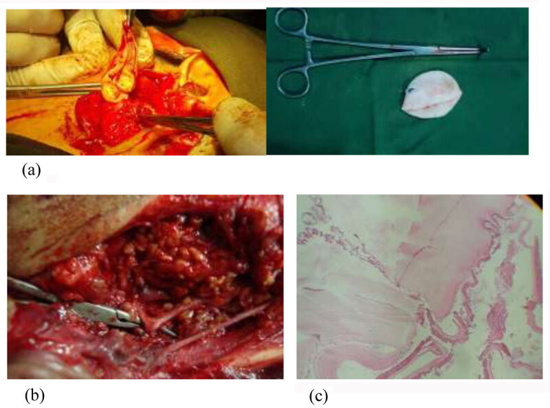
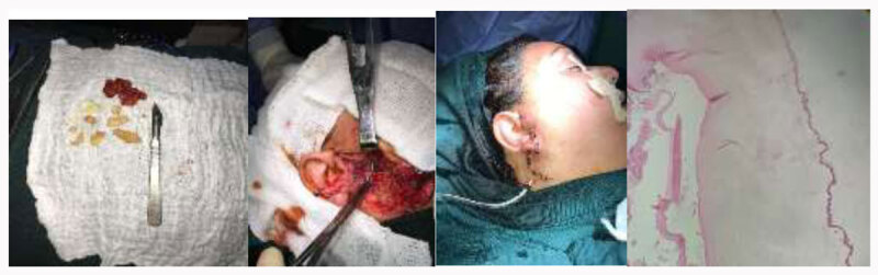
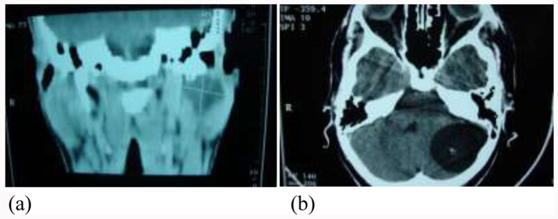
Hydatidosis (Echinococcosis) is a disease caused by infestation of hydatid cysts in any organ of body but mainly liver (70% of cases). Hydatidosis of salivary glands is rare and necessitate computerized tomography for diagnosis while fine needle aspiration remains controversial procedure.
Materials and methods: 6 patients diagnosed with hydatid cysts of parotid glands. These cases were admitted and treated at the maxillofacial surgery Clinic of the "AL-Ramadi" Hospital in Iraq. 5 patients were female and 1 male with age group was between 30-50 years. The patients complained of painless unilateral swelling in parotid region and who were diagnosed hydatid cysts using CT. All cases were treated by superficial parotidectomy with cystectomy and preservation of facial nerve.
Results: All hydatid cysts are CE1- type with no recurrences were reported in any of these cases. The postoperative edema was the most common complication. Other complications were not seen.
Conclusion: parotid hydatid cyst should be included in differential diagnosis of persistent parotid swelling especially those with history of hepatic hydatid cysts. Computerized tomography is the gold imaging that aid in diagnosis and classification of hydatid cysts. Most cases are CE1 type and Eosinophilia is a sign of concern in some patients. Surgical treatment remains the "gold standard" in therapy.
Highlights: Hydatidosis of parotid glands is rare but must be included in differential diagnosis of cystic swelling of salivary glands especially those with history of hepatic hydatid cysts.The hydatid cysts are classified according to morphology on imaging into 5 typesTotal serum bilirubin, eosinophilia and leukocytosis are seenSuperficial parotidectomy with removal of hydatid cysts is the treatment of choice in parotid hydatid cysts.
期刊介绍:
IJS Protocols is the first peer-reviewed, international, open access journal seeking to publish research protocols across across the full breadth of the surgical field. We are aim to provide rapid submission to decision times whilst maintaining a high quality peer-review process.


 分享
分享

 求助内容:
求助内容: 应助结果提醒方式:
 扫码关注我们
