IF 1.4 3区 医学Q4 BIOCHEMISTRY & MOLECULAR BIOLOGYMolecular VisionPub Date : 2021-11-19eCollection Date: 2021-01-01
Sara A Adelman, Kazuya Oikawa, Gopika Senthilkumar, Ralph Møller Trane, Leandro B C Teixeira, Gillian J McLellan
{"title":"在大眼青光眼模型中定位视网膜神经节细胞体。","authors":"Sara A Adelman, Kazuya Oikawa, Gopika Senthilkumar, Ralph Møller Trane, Leandro B C Teixeira, Gillian J McLellan","doi":"","DOIUrl":null,"url":null,"abstract":"<p><strong>Purpose: </strong>The purpose of this study was to identify a robust, representative region of interest (ROI) for studies of retinal ganglion cell (RGC) soma loss in feline congenital glaucoma (FCG), a spontaneous, large-eyed glaucoma model.</p><p><strong>Methods: </strong>Seven FCG and three wild-type (wt) eyes were collected from 10 adult cats of both sexes. Eyes enucleated postmortem were immediately fixed overnight in 4% paraformaldehyde and then stored in 0.1 M PBS at 4 °C. The retinas were wholemounted, Nissl stained with cresyl violet, and imaged using light microscopy. Somas of RGCs were manually identified according to long-established morphological criteria and quantified using a semiautomated method; their coordinates were used to create density maps and plots of the retinal topography. The RGC axon counts for the corresponding eyes were obtained from glutaraldehyde-fixed, resin-embedded optic nerve cross-sections stained with 0.1% p-phenylenediamine (PPD) using a semiautomated counting method. Correlations between total optic nerve axons and RGC soma counts were assessed by linear regression. A k-means cluster algorithm was used to identify a retinal ROI, with further definition using a probability density algorithm.</p><p><strong>Results: </strong>Interindividual variability in RGC total soma counts was more pronounced in FCG cats (mean = 83,244, range: 0-155,074) than in wt cats (mean = 117,045, range: 97,373-132,972). In general, RGC soma counts were lower in FCG cats than they were in wt cats. RGC axon counts in the optic nerve cross-sections were lower than, but strongly correlated to, the total RGC soma count across all cats (in wt and FCG retinas; R<sup>2</sup> = 0.88) and solely FCG eyes (R<sup>2</sup> = 0.92). The k-means cluster algorithm indicated a region of the greatest mean difference between the normal wt retinas and FCG-affected retinas within the temporal retina, incorporating the region of the area centralis.</p><p><strong>Conclusions: </strong>As in other species, RGC soma count and topography are heterogeneous between individual cats, but we identified an ROI in the temporal retina for future studies of RGC soma loss or preservation in a large-eyed model of congenital glaucoma. Many of the methods refined and established to facilitate studies in this FCG model will be broadly applicable to studies in other large-eyed models.</p>","PeriodicalId":18866,"journal":{"name":"Molecular Vision","volume":"27 ","pages":"608-621"},"PeriodicalIF":1.4000,"publicationDate":"2021-11-19","publicationTypes":"Journal Article","fieldsOfStudy":null,"isOpenAccess":false,"openAccessPdf":"https://ftp.ncbi.nlm.nih.gov/pub/pmc/oa_pdf/0c/92/mv-v27-608.PMC8645189.pdf","citationCount":"0","resultStr":"{\"title\":\"Mapping retinal ganglion cell somas in a large-eyed glaucoma model.\",\"authors\":\"Sara A Adelman, Kazuya Oikawa, Gopika Senthilkumar, Ralph Møller Trane, Leandro B C Teixeira, Gillian J McLellan\",\"doi\":\"\",\"DOIUrl\":null,\"url\":null,\"abstract\":\"<p><strong>Purpose: </strong>The purpose of this study was to identify a robust, representative region of interest (ROI) for studies of retinal ganglion cell (RGC) soma loss in feline congenital glaucoma (FCG), a spontaneous, large-eyed glaucoma model.</p><p><strong>Methods: </strong>Seven FCG and three wild-type (wt) eyes were collected from 10 adult cats of both sexes. Eyes enucleated postmortem were immediately fixed overnight in 4% paraformaldehyde and then stored in 0.1 M PBS at 4 °C. The retinas were wholemounted, Nissl stained with cresyl violet, and imaged using light microscopy. Somas of RGCs were manually identified according to long-established morphological criteria and quantified using a semiautomated method; their coordinates were used to create density maps and plots of the retinal topography. The RGC axon counts for the corresponding eyes were obtained from glutaraldehyde-fixed, resin-embedded optic nerve cross-sections stained with 0.1% p-phenylenediamine (PPD) using a semiautomated counting method. Correlations between total optic nerve axons and RGC soma counts were assessed by linear regression. A k-means cluster algorithm was used to identify a retinal ROI, with further definition using a probability density algorithm.</p><p><strong>Results: </strong>Interindividual variability in RGC total soma counts was more pronounced in FCG cats (mean = 83,244, range: 0-155,074) than in wt cats (mean = 117,045, range: 97,373-132,972). In general, RGC soma counts were lower in FCG cats than they were in wt cats. RGC axon counts in the optic nerve cross-sections were lower than, but strongly correlated to, the total RGC soma count across all cats (in wt and FCG retinas; R<sup>2</sup> = 0.88) and solely FCG eyes (R<sup>2</sup> = 0.92). The k-means cluster algorithm indicated a region of the greatest mean difference between the normal wt retinas and FCG-affected retinas within the temporal retina, incorporating the region of the area centralis.</p><p><strong>Conclusions: </strong>As in other species, RGC soma count and topography are heterogeneous between individual cats, but we identified an ROI in the temporal retina for future studies of RGC soma loss or preservation in a large-eyed model of congenital glaucoma. Many of the methods refined and established to facilitate studies in this FCG model will be broadly applicable to studies in other large-eyed models.</p>\",\"PeriodicalId\":18866,\"journal\":{\"name\":\"Molecular Vision\",\"volume\":\"27 \",\"pages\":\"608-621\"},\"PeriodicalIF\":1.4000,\"publicationDate\":\"2021-11-19\",\"publicationTypes\":\"Journal Article\",\"fieldsOfStudy\":null,\"isOpenAccess\":false,\"openAccessPdf\":\"https://ftp.ncbi.nlm.nih.gov/pub/pmc/oa_pdf/0c/92/mv-v27-608.PMC8645189.pdf\",\"citationCount\":\"0\",\"resultStr\":null,\"platform\":\"Semanticscholar\",\"paperid\":null,\"PeriodicalName\":\"Molecular Vision\",\"FirstCategoryId\":\"3\",\"ListUrlMain\":\"\",\"RegionNum\":3,\"RegionCategory\":\"医学\",\"ArticlePicture\":[],\"TitleCN\":null,\"AbstractTextCN\":null,\"PMCID\":null,\"EPubDate\":\"2021/1/1 0:00:00\",\"PubModel\":\"eCollection\",\"JCR\":\"Q4\",\"JCRName\":\"BIOCHEMISTRY & MOLECULAR BIOLOGY\",\"Score\":null,\"Total\":0}","platform":"Semanticscholar","paperid":null,"PeriodicalName":"Molecular Vision","FirstCategoryId":"3","ListUrlMain":"","RegionNum":3,"RegionCategory":"医学","ArticlePicture":[],"TitleCN":null,"AbstractTextCN":null,"PMCID":null,"EPubDate":"2021/1/1 0:00:00","PubModel":"eCollection","JCR":"Q4","JCRName":"BIOCHEMISTRY & MOLECULAR BIOLOGY","Score":null,"Total":0}
引用次数: 0
摘要
目的:本研究的目的是为猫先天性青光眼(FCG)(一种自发性大眼青光眼模型)的视网膜神经节细胞(RGC)体细胞丢失研究确定一个稳健的、有代表性的兴趣区(ROI)。方法:从10只雌雄猫身上采集7只FCG眼和3只野生型(wt)眼。死后去核的眼睛立即在4%多聚甲醛中固定过夜,然后在0.1 M PBS中保存,温度为4°C。整片视网膜,甲酚紫尼氏染色,光镜成像。根据长期建立的形态学标准,人工鉴定RGCs的体细胞,并使用半自动方法进行定量;他们的坐标被用来创建密度图和视网膜地形图。相应眼睛的RGC轴突计数来自戊二醛固定、树脂包埋的视神经横截面,用0.1%对苯二胺(PPD)染色,采用半自动计数方法。通过线性回归评估视神经轴突总数与RGC体细胞计数之间的相关性。使用k-均值聚类算法识别视网膜ROI,并使用概率密度算法进一步定义。结果:与wt猫(平均= 117,045,范围:97,373-132,972)相比,FCG猫(平均= 83,244,范围:0-155,074)的RGC总体细胞计数的个体间差异更为明显。一般来说,FCG猫的RGC体细胞计数低于wt猫。视神经横截面上的RGC轴突计数低于所有猫(wt和FCG视网膜)的RGC体细胞总数,但与之密切相关;R2 = 0.88)和单独FCG眼(R2 = 0.92)。k-means聚类算法指出了颞视网膜内正常wt视网膜与fcg影响视网膜之间平均差异最大的区域,包括中央区域区域。结论:与其他物种一样,个体猫的RGC体数量和地形是不一样的,但我们在颞视网膜中发现了一个ROI,为未来在大眼先天性青光眼模型中RGC体丢失或保存的研究提供了基础。为促进本FCG模型的研究而完善和建立的许多方法将广泛适用于其他大眼睛模型的研究。
Mapping retinal ganglion cell somas in a large-eyed glaucoma model.
Purpose: The purpose of this study was to identify a robust, representative region of interest (ROI) for studies of retinal ganglion cell (RGC) soma loss in feline congenital glaucoma (FCG), a spontaneous, large-eyed glaucoma model.
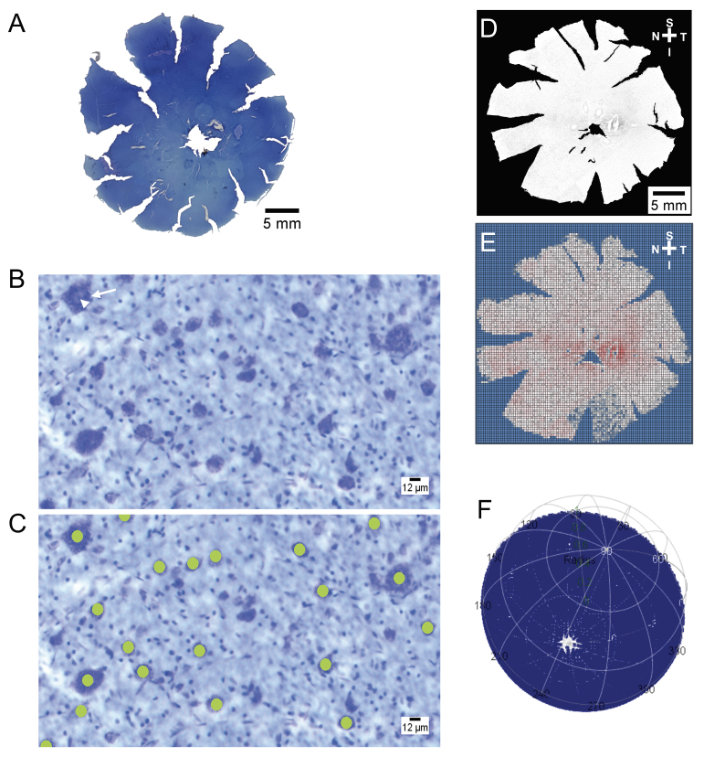
Methods: Seven FCG and three wild-type (wt) eyes were collected from 10 adult cats of both sexes. Eyes enucleated postmortem were immediately fixed overnight in 4% paraformaldehyde and then stored in 0.1 M PBS at 4 °C. The retinas were wholemounted, Nissl stained with cresyl violet, and imaged using light microscopy. Somas of RGCs were manually identified according to long-established morphological criteria and quantified using a semiautomated method; their coordinates were used to create density maps and plots of the retinal topography. The RGC axon counts for the corresponding eyes were obtained from glutaraldehyde-fixed, resin-embedded optic nerve cross-sections stained with 0.1% p-phenylenediamine (PPD) using a semiautomated counting method. Correlations between total optic nerve axons and RGC soma counts were assessed by linear regression. A k-means cluster algorithm was used to identify a retinal ROI, with further definition using a probability density algorithm.
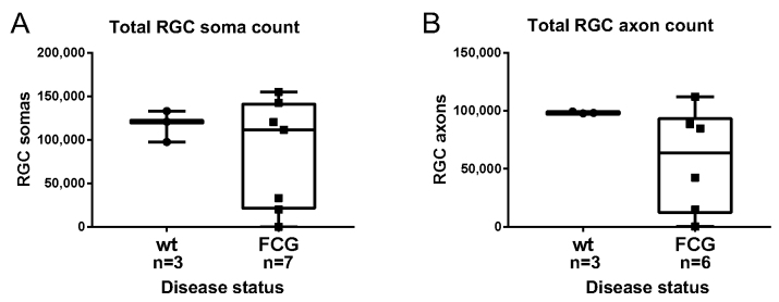
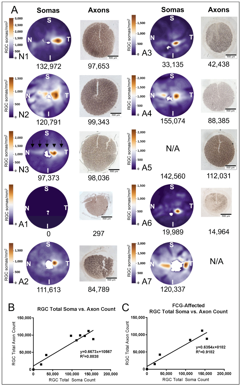
Results: Interindividual variability in RGC total soma counts was more pronounced in FCG cats (mean = 83,244, range: 0-155,074) than in wt cats (mean = 117,045, range: 97,373-132,972). In general, RGC soma counts were lower in FCG cats than they were in wt cats. RGC axon counts in the optic nerve cross-sections were lower than, but strongly correlated to, the total RGC soma count across all cats (in wt and FCG retinas; R2 = 0.88) and solely FCG eyes (R2 = 0.92). The k-means cluster algorithm indicated a region of the greatest mean difference between the normal wt retinas and FCG-affected retinas within the temporal retina, incorporating the region of the area centralis.
Conclusions: As in other species, RGC soma count and topography are heterogeneous between individual cats, but we identified an ROI in the temporal retina for future studies of RGC soma loss or preservation in a large-eyed model of congenital glaucoma. Many of the methods refined and established to facilitate studies in this FCG model will be broadly applicable to studies in other large-eyed models.
期刊介绍:
Molecular Vision is a peer-reviewed journal dedicated to the dissemination of research results in molecular biology, cell biology, and the genetics of the visual system (ocular and cortical).
Molecular Vision publishes articles presenting original research that has not previously been published and comprehensive articles reviewing the current status of a particular field or topic. Submissions to Molecular Vision are subjected to rigorous peer review. Molecular Vision does NOT publish preprints.
For authors, Molecular Vision provides a rapid means of communicating important results. Access to Molecular Vision is free and unrestricted, allowing the widest possible audience for your article. Digital publishing allows you to use color images freely (and without fees). Additionally, you may publish animations, sounds, or other supplementary information that clarifies or supports your article. Each of the authors of an article may also list an electronic mail address (which will be updated upon request) to give interested readers easy access to authors.
 分享
分享


 求助内容:
求助内容: 应助结果提醒方式:
 扫码关注我们
