Daria Kifjak, Johannes Leitner, Raphael Ambros, Benedikt H Heidinger, Ruxandra-Iulia Milos, Lucian Beer, Florian Prayer, Sebastian Röhrich, Helmut Prosch
{"title":"[弥漫性肺实质疾病的胸片表现]。","authors":"Daria Kifjak, Johannes Leitner, Raphael Ambros, Benedikt H Heidinger, Ruxandra-Iulia Milos, Lucian Beer, Florian Prayer, Sebastian Röhrich, Helmut Prosch","doi":"10.1007/s00117-021-00955-8","DOIUrl":null,"url":null,"abstract":"<p><strong>Clinical issue: </strong>Diffuse parenchymal lung diseases include a heterogeneous group of diseases of the lung parenchyma, the alveolar spaces, the vessels and the airways, which can be triggered by various pathomechanisms, such as inflammation and fibrotic changes. Since the therapeutic approaches and prognoses differ significantly between the diseases, the correct diagnosis is of fundamental importance. In routine clinical practice, next to the patients' history, the clinical presentation, the laboratory findings and the bronchoscopy, imaging plays a central role in establishing a diagnosis.</p><p><strong>Practical recommendations: </strong>The diagnosis of diffuse parenchymal lung diseases is an enormous challenge for clinicians, radiologists as well as pathologists and should therefore preferably be carried out in a multidisciplinary setting. Since patients often present with unspecific, respiratory symptoms, chest radiographs are the first imaging method used. Many patterns of diffuse parenchymal lung diseases (e.g., ground-glass opacities and consolidations), their distribution (e.g., cranial-caudal) and the presence of additional findings (e.g., mediastinal lymphadenopathy) are often already detectable on chest X‑rays. However, the imaging reference standard and thus, an integral part of the assessment of diffuse parenchymal lung disease, is the chest HR-CT. In some cases, the pattern of the HR-CT is pathognomonic, in others it is unspecific for a disease, so that further diagnostic steps are necessary.</p>","PeriodicalId":54513,"journal":{"name":"Radiologe","volume":"62 2","pages":"130-139"},"PeriodicalIF":0.0000,"publicationDate":"2022-02-01","publicationTypes":"Journal Article","fieldsOfStudy":null,"isOpenAccess":false,"openAccessPdf":"https://www.ncbi.nlm.nih.gov/pmc/articles/PMC8740870/pdf/","citationCount":"2","resultStr":"{\"title\":\"[Chest radiography findings in diffuse parenchymal lung diseases].\",\"authors\":\"Daria Kifjak, Johannes Leitner, Raphael Ambros, Benedikt H Heidinger, Ruxandra-Iulia Milos, Lucian Beer, Florian Prayer, Sebastian Röhrich, Helmut Prosch\",\"doi\":\"10.1007/s00117-021-00955-8\",\"DOIUrl\":null,\"url\":null,\"abstract\":\"<p><strong>Clinical issue: </strong>Diffuse parenchymal lung diseases include a heterogeneous group of diseases of the lung parenchyma, the alveolar spaces, the vessels and the airways, which can be triggered by various pathomechanisms, such as inflammation and fibrotic changes. Since the therapeutic approaches and prognoses differ significantly between the diseases, the correct diagnosis is of fundamental importance. In routine clinical practice, next to the patients' history, the clinical presentation, the laboratory findings and the bronchoscopy, imaging plays a central role in establishing a diagnosis.</p><p><strong>Practical recommendations: </strong>The diagnosis of diffuse parenchymal lung diseases is an enormous challenge for clinicians, radiologists as well as pathologists and should therefore preferably be carried out in a multidisciplinary setting. Since patients often present with unspecific, respiratory symptoms, chest radiographs are the first imaging method used. Many patterns of diffuse parenchymal lung diseases (e.g., ground-glass opacities and consolidations), their distribution (e.g., cranial-caudal) and the presence of additional findings (e.g., mediastinal lymphadenopathy) are often already detectable on chest X‑rays. However, the imaging reference standard and thus, an integral part of the assessment of diffuse parenchymal lung disease, is the chest HR-CT. In some cases, the pattern of the HR-CT is pathognomonic, in others it is unspecific for a disease, so that further diagnostic steps are necessary.</p>\",\"PeriodicalId\":54513,\"journal\":{\"name\":\"Radiologe\",\"volume\":\"62 2\",\"pages\":\"130-139\"},\"PeriodicalIF\":0.0000,\"publicationDate\":\"2022-02-01\",\"publicationTypes\":\"Journal Article\",\"fieldsOfStudy\":null,\"isOpenAccess\":false,\"openAccessPdf\":\"https://www.ncbi.nlm.nih.gov/pmc/articles/PMC8740870/pdf/\",\"citationCount\":\"2\",\"resultStr\":null,\"platform\":\"Semanticscholar\",\"paperid\":null,\"PeriodicalName\":\"Radiologe\",\"FirstCategoryId\":\"3\",\"ListUrlMain\":\"https://doi.org/10.1007/s00117-021-00955-8\",\"RegionNum\":4,\"RegionCategory\":\"医学\",\"ArticlePicture\":[],\"TitleCN\":null,\"AbstractTextCN\":null,\"PMCID\":null,\"EPubDate\":\"2022/1/7 0:00:00\",\"PubModel\":\"Epub\",\"JCR\":\"Q3\",\"JCRName\":\"Medicine\",\"Score\":null,\"Total\":0}","platform":"Semanticscholar","paperid":null,"PeriodicalName":"Radiologe","FirstCategoryId":"3","ListUrlMain":"https://doi.org/10.1007/s00117-021-00955-8","RegionNum":4,"RegionCategory":"医学","ArticlePicture":[],"TitleCN":null,"AbstractTextCN":null,"PMCID":null,"EPubDate":"2022/1/7 0:00:00","PubModel":"Epub","JCR":"Q3","JCRName":"Medicine","Score":null,"Total":0}
[Chest radiography findings in diffuse parenchymal lung diseases].
Clinical issue: Diffuse parenchymal lung diseases include a heterogeneous group of diseases of the lung parenchyma, the alveolar spaces, the vessels and the airways, which can be triggered by various pathomechanisms, such as inflammation and fibrotic changes. Since the therapeutic approaches and prognoses differ significantly between the diseases, the correct diagnosis is of fundamental importance. In routine clinical practice, next to the patients' history, the clinical presentation, the laboratory findings and the bronchoscopy, imaging plays a central role in establishing a diagnosis.
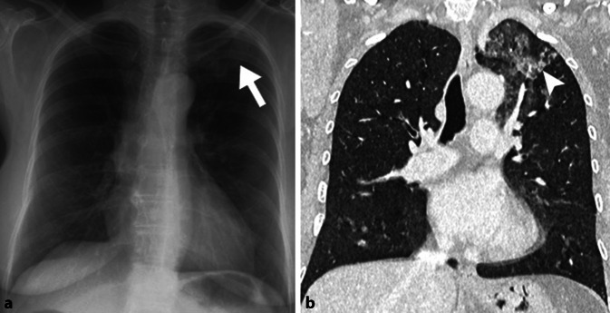
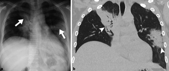
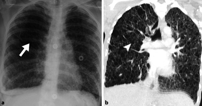
Practical recommendations: The diagnosis of diffuse parenchymal lung diseases is an enormous challenge for clinicians, radiologists as well as pathologists and should therefore preferably be carried out in a multidisciplinary setting. Since patients often present with unspecific, respiratory symptoms, chest radiographs are the first imaging method used. Many patterns of diffuse parenchymal lung diseases (e.g., ground-glass opacities and consolidations), their distribution (e.g., cranial-caudal) and the presence of additional findings (e.g., mediastinal lymphadenopathy) are often already detectable on chest X‑rays. However, the imaging reference standard and thus, an integral part of the assessment of diffuse parenchymal lung disease, is the chest HR-CT. In some cases, the pattern of the HR-CT is pathognomonic, in others it is unspecific for a disease, so that further diagnostic steps are necessary.
期刊介绍:
Der Radiologe is an internationally recognized journal dealing with all aspects of radiology and serving the continuing medical education of radiologists in clinical and practical environments. The focus is on x-ray diagnostics, angiography computer tomography, interventional radiology, magnet resonance tomography, digital picture processing, radio oncology and nuclear medicine.
Comprehensive reviews on a specific topical issue focus on providing evidenced based information on diagnostics and therapy.
Freely submitted original papers allow the presentation of important clinical studies and serve the scientific exchange.
Review articles under the rubric ''Continuing Medical Education'' present verified results of scientific research and their integration into daily practice.


 分享
分享
 求助内容:
求助内容: 应助结果提醒方式:
 扫码关注我们
